PET imaging with F-18 sodium fluoride (NaF) can detect activity related to tears in the outer wall of the aorta and could help manage patients with acute aortic syndrome, according to a study published March 16 in JACC: Cardiovascular Imaging.
U.K. researchers at the British Heart Foundation (BHF) in Edinburgh, Scotland, investigated whether F-18 NaF-PET/CT scans could show damaged aortas in patients with acute aortic syndrome. They found patients who had increased uptake of F-18 NaF were at higher risk of major clinical events, including death.
"We suggest that F-18 NaF PET holds promise as a marker of disease activity in acute aortic syndrome and may predict future clinical outcome and guide patient management," wrote lead investigator Dr. David Newby, a professor of cardiology at British Heart Foundation.
Acute aortic syndrome consists of a range of potentially catastrophic conditions, such as aortic dissections, which are tears in the inner layer of the aorta due to hypertension or atherosclerosis. Initial treatments typically involve the rapid control of blood pressure or surgery to repair the tear.
Diagnosing acute aortic syndrome in the clinic can be performed using CT, echocardiography, and MRI, yet PET may have an advantage over these anatomical imaging methods by detecting early molecular processes involved in the disease, according to the authors.
In previous work, Newby and colleagues have shown that F-18 NaF PET/CT scans offer a novel approach for quantifying vascular injury due to early plaque development (microcalcification) in patients with atherosclerosis.
In this study, the group aimed to determine if F-18 NaF PET/CT scans could reveal disease activity in tears in the aortic wall, and whether the findings could predict outcomes for patients.
The group enrolled 63 patients over the age of 25 with newly diagnosed but clinically stable acute aortic syndrome (aortic dissection or intramural hematomas). Patients were enrolled over a 24-month recruitment period that ended in November, 2021 and underwent F-18 NaF-PET/CT scans (Biograph mCT, Siemens Healthineers) within 12 weeks of their diagnosis.
The researchers measured aortic F-18 NaF uptake at the most diseased segment of the aorta based on the maximum tissue-to-background ratio (TBRmax). Radiotracer uptake was compared with changes in aortic size and major adverse aortic events, such as rupture or aorta-related death.
The mean follow-up duration from F-18 NaF PET/CT scan was 14.6 months. During this period, 12 patients experienced major adverse aortic events. Of these, two patients experienced aortic rupture and subsequent death. Open surgical repair was necessary in eight patients, and two patients required delayed endovascular stenting.
Compared with clinically normal patients, the researchers observed patients with acute aortic syndrome had increased F-18 NaF uptake (TBRmax: 1.36 +/- 0.39 vs. 2.02 +/- 0.42) with enhanced uptake at the site of disruptions in the inner aortic lining (+27.5%; p < 0.001).
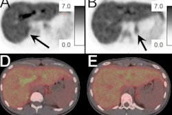
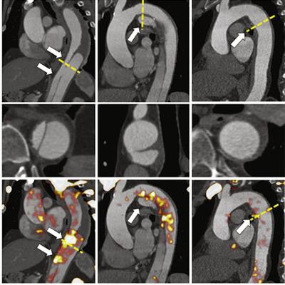
 (Top) Longitudinal and (bottom) cross-sectional (yellow dashed line) aortic imaging at sites of dissection and hematoma (white arrows). Image courtesy of JACC: Cardiovascular Imaging.
In addition, F-18 NaF uptake in the false lumen was associated with aortic growth (+7.1 mm/year; p = 0.011), and uptake of the tracer in the outer aortic wall was associated with major adverse aortic events (HR: 8.5; p = 0.019).
"In patients with acute aortic syndrome, F-18 NaF uptake was enhanced at sites of disease activity and was associated with aortic growth and clinical events," the researchers wrote.
Ultimately, this was a proof-of-concept study that demonstrated a potential application of F-18 NaF-PET/CT in patients with acute aortic syndrome. The researchers plan additional investigations to determine if the approach offers a predictive advantage over traditional clinical risk factors and anatomical imaging.
"Future studies in larger homogenous cohorts are now required to establish the clinical usefulness of F-18 NaF-PET/CT in patients with acute aortic syndrome," Newby and colleagues concluded.