Although 73% of chest x-rays show abnormalities in children with recurrent wheezing, they rarely lead to changes in diagnosis or treatment, supporting selective rather than routine imaging in this population.
- 73% of chest x-rays in children under five with recurrent wheezing showed abnormal findings, most commonly peribronchial cuffing (35%)
- Despite high abnormality rates, only 1% triggered further diagnostic workup and 0% led to changes in therapy or diagnosis
- Current clinical guidelines do not recommend routine chest radiography in children with recurrent wheezing absent atypical or severe features
- Children with elevated blood eosinophil counts (≥150 cells/µL) had significantly higher rates of abnormal x-rays (92% versus 71%)
- Study recommends clinically driven rather than routine use of chest x-rays to minimize unnecessary radiation exposure and procedural stress in young children
Abnormal chest x-rays are frequent in preschool children with recurrent wheezing, but rarely lead to changes in diagnosis or clinical management, according to a recent study.
The finding is from a retrospective analysis of 271 children under five years of age who presented with recurrent wheezing at a pediatric pulmonology outpatient clinic, and adds real-world evidence supporting selective rather than routine use of chest x-rays in preschoolers, according to the authors.
“Given the risks associated with radiation exposure in young children and procedural stress, it is crucial to better understand the true diagnostic value of [chest x-rays] in this population,” noted lead author Emma Lück, MD, of the University Hospital of Cologne in Germany, and colleagues.
Recurrent wheezing affects roughly half of all children under three years old, and although current clinical guidelines do not recommend routine chest radiography in the absence of atypical or severe features, clinicians still commonly order chest x-rays to rule out pulmonary abnormalities, the authors explained.
To investigate whether practice offers real diagnostic value, the researchers conducted a retrospective chart review of children under five years old who presented with recurrent wheezing to the outpatient clinic and had undergone a chest x-ray within four weeks of their visit. Children with an acute respiratory infection at the time of imaging or pre-existing pulmonary or cardiac disease were excluded. The team recorded demographic and clinical characteristics, laboratory values including blood eosinophil counts, and whether chest x-ray findings led to further diagnostic workup, a change in therapy, or a revised diagnosis.
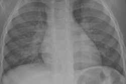
According to the analysis, chest x-rays were read as abnormal in 73% of cases, most often showing peribronchial cuffing (35%), followed by peribronchial opacities, parenchymal consolidation, and accentuated bronchial markings (13% each), and pulmonary hypertransparency (11%).
However, despite this high rate of abnormal findings, further diagnostic workup was triggered in just three cases (1%), and no chest x-ray result led to a change in therapy or diagnosis, the researchers reported.
In secondary results, children with elevated blood eosinophil counts (≥ 150 cells/µL) had significantly higher rates of abnormal chest x-rays than those with lower counts (92% versus 71%; odds ratio, 4.43; p = 0.023). Age, sex, prematurity, and personal or parental atopic history were not associated with abnormal findings, according to the findings.
Ultimately, the findings are consistent with current clinical guidelines on recurrent wheezing, which do not consider chest x-rays an essential diagnostic tool in this setting, the authors noted.
“Our findings are consistent with current recommendations advocating a clinically driven rather than routine use of chest radiography in preschool children with recurrent wheezing,” the researchers concluded.
The study was published July 15 in BMC Pediatrics.