An AI model called OccuNet achieved 97.5% sensitivity in detecting femoral-neck fractures on radiographs, outperforming radiologists and emergency medicine physicians, particularly for fractures that initially appear negative or unclear on x-rays. The AI tool not only improved diagnostic accuracy but also reduced reading times by up to 19%.
- OccuNet achieved 97.5% sensitivity and 98.8% specificity in detecting femoral-neck fractures across pooled test groups
- For radiograph-negative or indeterminate fractures, OccuNet's sensitivity was 94.7%, significantly higher than radiologists (86.2%) and emergency physicians (68.8%)
- AI assistance improved radiologist sensitivity from 93.7% to 97.2% and emergency physician sensitivity from 84.3% to 95.6%
- Reading times decreased by 14.9% for radiologists and 18.9% for emergency physicians when using AI assistance
An AI model performed well in detecting femoral-neck fractures on radiographs, according to findings published September 8 in Radiology.
An AI tool called OccuNet bested radiologists and emergency medicine physicians in finding these fractures on pelvic or hip radiographs, wrote a group of researchers led by Nai-Feng Tian, MD, PhD, from The Second Affiliated Hospital and Yuying Children’s Hospital of Wenzhou Medical University in Wenzhou, China.
“These results support prospective evaluation of AI-assisted clinical workflow integration,” Tian and colleagues wrote.
Prior reports suggest that about 40% of patients with negative or equivocal radiographs are later diagnosed at CT or MRI. For these radiograph-negative or indeterminate femoral-neck fractures, initial radiographs showed no definite fracture. But same-episode CT or MRI confirmed Garden I/II fractures.
Routine CT or MRI for these patients can increase cost and prolong emergency department stays. AI in previous studies has demonstrated high levels of accuracy for radiographic hip-fracture detection, comparable to that of experts.
Tian and co-authors developed and externally evaluated OccuNet for finding femoral-neck fractures on pelvic or hip radiographs. They also compared OccuNet’s performance and accuracy with those of radiologists and emergency medicine physicians and evaluated the tool’s effect on reader performance and accuracy.
The study included 2,576 patients suspected of having hip trauma who underwent pelvic or hip radiography and same-episode CT or MRI at four hospitals between 2009 and 2025. Patient data were placed into a training set (n = 810), an internal test set (n = 760), and three external test sets (total n = 1,006).
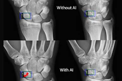
 Example anteroposterior (AP) pelvic radiographs showing simulated acquisition artifacts used for stage 1 artifact-robust pretraining. Images are derived from the same AP pelvic radiograph from a 75-year-old woman with a CT- or MRI-confirmed left femoral-neck fracture. (A) Original AP pelvic radiograph. (B) Simulated grid or cable-shadow artifact. (C) Simulated motion-blur artifact. (D) Simulated sensor saturation or overexposure artifact.RSNA
In the pooled group (n = 1,766), the model achieved 97.5% sensitivity, 98.8% specificity, and an area under the curve (AUC) of 0.99.
For radiograph-negative or indeterminate fractures (n = 189), the model achieved a higher sensitivity (94.7%) than the radiologists (86.2%, p < 0.001) and emergency medicine physicians (68.8%, p < 0.001).
AI help with OccuNet led to improved sensitivities for both professional groups. This included increases from 93.7% to 97.2% for radiologists (p < 0.001) and from 84.3% to 95.6% for emergency medicine physicians (p < 0.001).
Finally, the researchers reported that OccuNet led to average reading times being shortened by 14.9% for radiologists and by 18.9% for the emergency medicine physicians (both p < 0.001).
The researchers called for prospective workflow studies to evaluate AI-first triage, calibrated confidence displays, and second-look prompts for high-confidence AI-positive radiographs.
These sentiments were shared in an accompanying editorial written by Dyan Flores, MD, and Tatiane Cantarelli, MD, from the University of Ottawa and The Ottawa Hospital in Canada. These prospective studies should compare diagnostic pathways incorporating AI-assisted radiography, CT, and MRI. These approaches will decide whether AI improves patient selection for advanced imaging while supporting diagnostic safety, the editorial authors wrote.
“Beyond diagnostic accuracy, implementation studies should evaluate how AI integrates into clinical workflows, including its effects on healthcare resource utilization, emergency department throughput, downstream imaging, patient outcomes, and overall costs,” they wrote.
Read the full study here.