X-ray imaging reveals that nearly two in five patients undergoing maintenance hemodialysis have severe abdominal aortic calcification, a group in Palestine has reported.
Researchers from An-Najah National University Hospital in Nablus assessed abdominal aortic calcification burden and its predictors in a local hemodialysis cohort, with findings suggesting that lateral lumbar x-ray imaging may serve as an accessible cardiovascular risk stratification tool, according to the researchers.
“These findings support the value of AAC as a clinically relevant vascular phenotype in dialysis populations and suggest that simple lateral lumbar radiography may be useful for opportunistic risk stratification in appropriate settings,” the group wrote. The study was published September 19 in Scientific Reports.
Cardiovascular disease is the leading cause of death in patients with chronic kidney disease on maintenance hemodialysis, with abdominal aortic calcification recognized as an independent cardiovascular risk marker in this population, the authors wrote. Although studies in Western, European, Asian, and African hemodialysis cohorts have reported high prevalence, data from Palestinian and Middle Eastern populations remain scarce, they added.
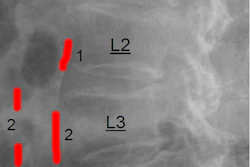
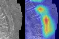
To bridge the gap, the group recruited 163 hemodialysis patients at their hospital between August 2025 and January 2026. Patients underwent lateral lumbar x-rays scored with a validated 24-point semi-quantitative system (Kauppila method) that rates aortic wall calcification at four lumbar vertebral levels: categorized as no calcification (0), mild-to-moderate (1 to 6), or severe (7 or higher).
According to the findings, lateral lumbar radiographs scored using the Kauppila method revealed a high burden of aortic calcification across the cohort: 35.6% of patients (median age, 50.5 years old) showed no calcification, 23.9% (median age, 61 years old) scored in the mild-to-moderate range, and 40.5% (median age, 66 years old) met the threshold for severe calcification. Radiographic severity tracked closely with age, with a Spearman correlation of ρ = 0.531 (p<0.001).
In multivariable analysis, age was the only factor independently and consistently associated with a worse radiographic classification, with each additional year of age tied to a 7.9% higher odds of belonging to a more severe calcification category (adjusted OR 1.079, p < 0.001), the researchers reported.
“Abdominal aortic calcification was common among Palestinian patients receiving maintenance hemodialysis, with severe calcification affecting approximately two in five participants,” the group wrote.
The findings reinforce lateral lumbar radiography's potential as an accessible, low-cost tool for opportunistic cardiovascular risk stratification in dialysis care, the authors suggested, noting that the current data support risk awareness rather than universal screening recommendations.
Prospective multicenter studies with larger samples, standardized imaging reproducibility, and longitudinal outcome assessment are needed to clarify whether modifiable pathways beyond age meaningfully influence AAC burden and progression, the researchers concluded.
The full study is available here.