A U.S. Food and Drug Administration (FDA)-cleared AI tool shows 99.2% sensitivity for pulmonary embolism (PE) detection when used with radiologist readers -- outperforming the algorithm operating alone, researchers have reported.
The findings highlight the continued central role of radiologists, noted a team led by Shlomit Goldberg-Stein, MD, of Northwell Health in New Hyde Park, NY. The group included researchers from the Harvey L. Neiman Health Policy Institute (HPI), and study results were published May 13 in Radiology: Artificial Intelligence.
"We observed a high … agreement between radiologists and the AI tool for the diagnosis of PE, with both AI and AI-informed radiologists offering unique diagnostic contributions," the authors wrote.
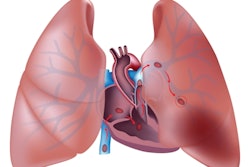
PE is the third most common cause of cardiovascular disease-related deaths, they explained, writing that the current standard of care for detecting PET is CT pulmonary angiography (CTPA). The exam is effective, but interpretations can be delayed due to increased radiologist workloads and shortages. That's why using AI to read these studies could expedite care, according to the group.
Goldberg-Stein and colleagues investigated the use of an AI tool for this indication (Aidoc, Tel Aviv, Israel) via a study that included data from 32,501 CTPA examinations performed in 29,492 patients at Northwell Health between August 2021 and February 2023.
They reported the following:
- Overall agreement between the algorithm and interpreting radiologists was 97.8%. This agreement was higher for AI-negative than AI-positive exams (98.2% versus 93.8%), suggesting "AI-negative outputs provide supportive signal while AI-positive alerts merit scrutiny."
- Among the 9.9% of exams that were PE-positive, 84% were detected by both the radiologist and AI working together, while 15% were identified by radiologists alone. Only 0.8% were caught exclusively by the AI tool.
- Reconciliation of disagreements confirmed the radiologist's interpretation in 88.7% of discordant cases.
Agreement varied by PE characteristics, with higher concordance for acute compared to chronic emboli (87.3% versus 60.1%) and higher concordance for central PE than for peripheral PE (central, 95.8%, and lobar/segmental, 83.8%) -- a result "consistent with the tool's triage focus."
 Case example of a false-negative AI result. An 85-year-old female presented to the emergency department with dyspnea. Axial 0.625-mm from CT pulmonary angiography examination with contrast (CTPA) images demonstrate a thin, linear right-sided filling defect extending from the right interlobar pulmonary artery (A and B, red arrows) into the right middle lobar artery (B, yellow arrow), consistent with acute pulmonary embolism (PE). The finding was described in the radiology report; however, the AI result was negative for PE. Adjudication was consistent with acute PE.Radiology: Artificial Intelligence

"AI-informed radiologists achieved a sensitivity of 99.2% for pulmonary embolism detection," Goldberg-Stein said in a statement released by HPI. "Radiologist-AI agreement was highest for acute and central emboli -- the cases associated with the greatest clinical urgency and mortality risk. This suggests the algorithm is most reliable in precisely the clinical scenarios where triage has the greatest potential to impact patient outcomes."
Access the full study here.