
A quarter of patients hospitalized with COVID-19 show pulmonary embolism (PE) on CT pulmonary angiography (CTPA) or nuclear stress testing, according to a study published July 13 in Radiology.
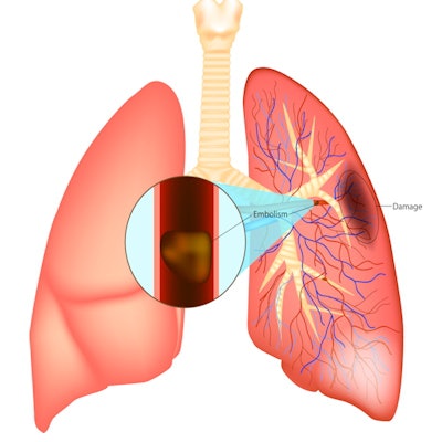
The condition is caused by blocked arteries in the lung, and if untreated, it has a mortality rate of up to 30%, according to a statement released by the RSNA.
A team led by Dr. Sadjad Riyahi of Weill Cornell Medicine in New York City used electronic medical record data from 413 people hospitalized with COVID-19 between March 3 and June 5, 2020. Of these patients, 102 were diagnosed with pulmonary embolism via chest CT or stress testing, and the rate of acute PE was greater in men by 74% and in smokers by 86%. The rate of acute pulmonary embolism did not differ significantly between intensive care and nonintensive care patients, however (29% compared to 24%).
Questions about COVID-19-induced pulmonary embolism and how best to diagnose it remain, wrote Dr. Loren Ketai of the University of New Mexico in Albuquerque in an accompanying editorial.
"While this study confirms several important clinical observations regarding thromboembolism and COVID-19, key practical questions remain unanswered. Not the least important, is how to best identify patients with COVID-19 who will benefit from pulmonary CTPA. ... Even when pulmonary emboli are detected by CTPA or perfusion scintigraphy, their clinical significance in the setting of COVID-19 may be ambiguous," he wrote.


![Axial images from unenhanced calcium score cardiac CT (left) and curved planar reformation images from CT angiography (right) show that higher long-term exposure to air pollution is associated with greater coronary artery calcium and more obstructive coronary artery disease (CAD). Top row: Images in a 68-year-old male patient with higher 10-year mean ambient air pollution exposure (7.9 μg/m3 for particulate matter measuring ≤2.5 μm in diameter [PM2.5] and 17.4 parts per billion [ppb] for NO2) with extensive CAD (coronary artery calcium score [CACS] >1,000 and obstructive CAD [≥70% diameter stenosis]). Bottom row: Images in a 57-year-old female patient with lower 10-year mean ambient air pollution exposure (6.3 μg/m3 for PM2.5 and 4.6 ppb for NO2) with no CAD (CACS = 0 and no obstructive stenosis).](https://img.auntminnie.com/mindful/smg/workspaces/default/uploads/2026/06/hanneman.r6SMLzkezo.png?auto=format%2Ccompress&fit=crop&h=100&q=70&w=100)


![Axial images from unenhanced calcium score cardiac CT (left) and curved planar reformation images from CT angiography (right) show that higher long-term exposure to air pollution is associated with greater coronary artery calcium and more obstructive coronary artery disease (CAD). Top row: Images in a 68-year-old male patient with higher 10-year mean ambient air pollution exposure (7.9 μg/m3 for particulate matter measuring ≤2.5 μm in diameter [PM2.5] and 17.4 parts per billion [ppb] for NO2) with extensive CAD (coronary artery calcium score [CACS] >1,000 and obstructive CAD [≥70% diameter stenosis]). Bottom row: Images in a 57-year-old female patient with lower 10-year mean ambient air pollution exposure (6.3 μg/m3 for PM2.5 and 4.6 ppb for NO2) with no CAD (CACS = 0 and no obstructive stenosis).](https://img.auntminnie.com/mindful/smg/workspaces/default/uploads/2026/06/hanneman.r6SMLzkezo.png?auto=format%2Ccompress&fit=crop&h=112&q=70&w=112)


