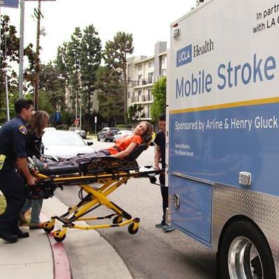
In a pilot project, University of California, Los Angeles (UCLA) Health has launched a specialized ambulance unit that is prepared to treat stroke patients on the spot using a mobile CT scanner.
As part of the pilot program, the ambulance began responding to emergency calls in Santa Monica, CA, this September. The stroke unit has a CT scanner (CereTom, Samsung NeuroLogica) and also a mobile blood-testing laboratory. A neurologist, radiologic technologist, critical care nurse, and paramedic ride inside the vehicle.
 Image courtesy of UCLA Health.
Positive feedback has encouraged the program organizers to consider expanding the effort to a fleet of four to nine units that would operate throughout Los Angeles County. In addition, the Los Angeles County Board of Supervisors recently voted to endow the program with nearly $1.5 million in additional funding to allow the vehicle to offer services on a weekly basis for up to 30 months.
Previous research indicates that for every minute that goes by without treatment in a typical stroke, 2 million brain cells die, UCLA noted. The program's goal is to enable treatment to start earlier than if patients had to be transported to a hospital.
"With the UCLA Health Mobile Stroke Unit, we are bringing the hospital to the patient instead of the patient to the hospital, in order to save as much brain as possible," said Dr. Jeffrey Saver, director of the UCLA Comprehensive Stroke Center, in a release from the university.
There are approximately 14 mobile stroke units in operation or under development in the U.S., according to UCLA.