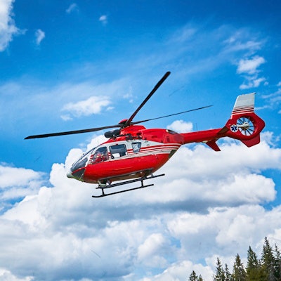
Putting a point-of-care ultrasound (POCUS) scanner aboard an emergency helicopter can help medical personnel triage trauma cases for patients in rural areas that are distant from tertiary care hospitals, according to a study by Canadian researchers presented at the recent meeting of the American College of Emergency Physicians (ACEP).
Because the largely rural landscape of Canada and its dispersed population makes it a challenge to rapidly respond to traumas that occur at long distances from tertiary centers, helicopter emergency medical services take action to manage these situations, explained Dr. Tom Jelic of the University of Manitoba in Winnipeg.
But it can be challenging for emergency medical personnel to make assessments in the helicopter using their clinical judgement, explained Jelic, who is point-of-care ultrasound lead for Shock Trauma Air Rescue Service (STARS) in Manitoba,
"With the vibrations of the helicopter, your ears are essentially useless," he said in an interview with AuntMinnie.com. "There is also glare that makes it difficult to do an appropriate physical examination. With the ultrasound machine, it can help you to determine if the patient has a collapsed lung or has blood in the chest or in the abdomen."
Moreover, in trauma medicine, quick, qualitative assessment of a patient is often needed to avoid grave outcomes like death, according to Jelic. To this end, air medical crew use focused assessment with sonography in trauma (FAST) and extended FAST (eFAST) exams to rapidly evaluate trauma patients. Information provided from POCUS is relayed to the tertiary center, where a trauma surgeon, for example, can prepare for surgery or another procedure, he said.
In their paper presented at ACEP 2020, Jelic and co-investigators sought to investigate the efficacy of POCUS performed on helicopters used for the STARS missions. They evaluated data on POCUS images, interpretations, and clinical integration over a two-year period and compared it with the gold standard -- CT or operative reports. They also looked at whether the use of POCUS delayed time at the scene.
A total of 231 patients were transported by helicopter and had an ultrasound performed while 1,016 were transported without an ultrasound performed. The mean scene time was no different for patients who underwent ultrasound compared with those who did not: 25.98 minutes (+/- 3.18, 95% CI) compared with 26.99 minutes (+/- 1.44, 95% CI), yielding a difference that was not significant (p < 0.1336).
The result translates to the fact that ultrasound performed by the air medical crew did not prolong the time they were at the scene according to Jelic.
The sensitivity and specificity numbers, denoting the accuracy of POCUS compared with CT or operative report findings, were 52.5 and 89.9, respectively, in the study.
"We have a pretty robust quality assurance program where the flight crew is required to store and save the images on the server," Jelic said. "I review the images and the interpretation of what [the air medical crew] thought the image showed."
Scans that were false positives or negatives did not adversely affect patient care or outcomes, Jelic added.
Extremely cold weather (-40 degrees Celsius [-40 degrees Fahrenheit] and below) can be a factor that limits the immediate use of POCUS in emergency situations, noted Dr. Jelic.
"We haven't had many experiences where we have had issues with it (POCUS)," he said. "When it's very cold, the screen can sometimes be slow to respond."



![Examples of ultrasound findings and techniques. (A) Images in a 39-year-old male patient with a mass in the left thigh. The mass is heterogeneous on the B-mode US image (compared with the patient in D) and showed increased microvascularity (superb microvascular imaging [SMI]) and shear-wave elastography (SWE) values. Undifferentiated pleomorphic sarcoma was diagnosed at biopsy (with pleomorphic rhabdomyosarcoma in surgical specimen). (B) Images in an 18-year-old male patient with a mass in the left leg. The mass is hypoechoic on the B-mode image, with no other findings suggestive of malignancy. The lesion is in contact with the cortex of the tibia, which is slightly irregular. CT revealed a doubtful anteromedial tibial erosion. The microvascular study demonstrated high vascularization, suggestive of malignancy. Periosteal Ewing sarcoma was diagnosed with both histologic and immunohistochemical confirmation. (C) Images in a 69-year-old female patient with a lump growing on the outside of the left leg. Multiple SWE examinations were performed (please note the high values obtained in the measurements, whereas the color map highlights the stiffness relative to adjacent tissues). SMI showed areas of increased vascularization to target for sampling. Undifferentiated spindle cell sarcoma was diagnosed at biopsy, with residual leiomyosarcoma in the surgical specimen after neoadjuvant therapy. (D) Images in a 56-year-old female patient with a mass in the right thigh. The mass is heterogeneous at both B-mode ultrasound (similar to patient A) and MRI (coronal T2-weighted spectral attenuated inversion recovery [SPAIR]; T1-weighted pre-contrast and postcontrast imaging), which even shows uptake after the administration of paramagnetic contrast material, which is traditionally suggestive of malignancy. Low values at SMI and elastography are suggestive of benignity. Spindle cell lipoma was diagnosed at biopsy, with atypical spindle cell lipomatous tumor in the surgical specimen.](https://img.auntminnie.com/mindful/smg/workspaces/default/uploads/2026/08/images-radiol250278fig2.APCFLSvX6p.jpg?auto=format%2Ccompress&fit=crop&h=100&q=70&w=100)


![Examples of ultrasound findings and techniques. (A) Images in a 39-year-old male patient with a mass in the left thigh. The mass is heterogeneous on the B-mode US image (compared with the patient in D) and showed increased microvascularity (superb microvascular imaging [SMI]) and shear-wave elastography (SWE) values. Undifferentiated pleomorphic sarcoma was diagnosed at biopsy (with pleomorphic rhabdomyosarcoma in surgical specimen). (B) Images in an 18-year-old male patient with a mass in the left leg. The mass is hypoechoic on the B-mode image, with no other findings suggestive of malignancy. The lesion is in contact with the cortex of the tibia, which is slightly irregular. CT revealed a doubtful anteromedial tibial erosion. The microvascular study demonstrated high vascularization, suggestive of malignancy. Periosteal Ewing sarcoma was diagnosed with both histologic and immunohistochemical confirmation. (C) Images in a 69-year-old female patient with a lump growing on the outside of the left leg. Multiple SWE examinations were performed (please note the high values obtained in the measurements, whereas the color map highlights the stiffness relative to adjacent tissues). SMI showed areas of increased vascularization to target for sampling. Undifferentiated spindle cell sarcoma was diagnosed at biopsy, with residual leiomyosarcoma in the surgical specimen after neoadjuvant therapy. (D) Images in a 56-year-old female patient with a mass in the right thigh. The mass is heterogeneous at both B-mode ultrasound (similar to patient A) and MRI (coronal T2-weighted spectral attenuated inversion recovery [SPAIR]; T1-weighted pre-contrast and postcontrast imaging), which even shows uptake after the administration of paramagnetic contrast material, which is traditionally suggestive of malignancy. Low values at SMI and elastography are suggestive of benignity. Spindle cell lipoma was diagnosed at biopsy, with atypical spindle cell lipomatous tumor in the surgical specimen.](https://img.auntminnie.com/mindful/smg/workspaces/default/uploads/2026/08/images-radiol250278fig2.APCFLSvX6p.jpg?auto=format%2Ccompress&fit=crop&h=112&q=70&w=112)