A new class of molecular mutation seen in various forms of breast cancer has been identified, according to a study published in the April 15 issue of Cancer Research. Identification of the mutation, called fusion transcripts, by Mayo Clinic researchers may provide a way to identify tumor subtypes and lead to new strategies to treat them.
The study was undertaken to systematically search for fusion genes and fusion transcripts linked to different types of breast tumors. Fusion transcripts are created when chromosomes break apart and recombine, an event that commonly occurs in cancer cells.
Fusion genes are created during this process when two halves of normal genes become linked. Fusion genes (DNA) create fusion transcripts (RNA), which then produce fusion proteins (Cancer Research, April 15, 2012, Vol. 72:8, pp. 1921-1928).
The research findings indicate that fusion transcripts are much more common in breast cancer than previously thought, said senior investigator Dr. Edith Perez, deputy director of the Mayo Clinic Cancer Center in Jacksonville, FL. The fusion transcripts represent a new class of mutation with an unknown role in breast cancer.
The finding that some fusion transcripts are tumor subtype-specific suggests that these entities may be critical determinants in the etiology of breast cancer subtypes, useful as biomarkers for tumor stratification, or exploitable as cancer-specific therapeutic targets, according to the authors.


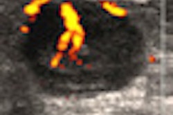


![Examples of ultrasound findings and techniques. (A) Images in a 39-year-old male patient with a mass in the left thigh. The mass is heterogeneous on the B-mode US image (compared with the patient in D) and showed increased microvascularity (superb microvascular imaging [SMI]) and shear-wave elastography (SWE) values. Undifferentiated pleomorphic sarcoma was diagnosed at biopsy (with pleomorphic rhabdomyosarcoma in surgical specimen). (B) Images in an 18-year-old male patient with a mass in the left leg. The mass is hypoechoic on the B-mode image, with no other findings suggestive of malignancy. The lesion is in contact with the cortex of the tibia, which is slightly irregular. CT revealed a doubtful anteromedial tibial erosion. The microvascular study demonstrated high vascularization, suggestive of malignancy. Periosteal Ewing sarcoma was diagnosed with both histologic and immunohistochemical confirmation. (C) Images in a 69-year-old female patient with a lump growing on the outside of the left leg. Multiple SWE examinations were performed (please note the high values obtained in the measurements, whereas the color map highlights the stiffness relative to adjacent tissues). SMI showed areas of increased vascularization to target for sampling. Undifferentiated spindle cell sarcoma was diagnosed at biopsy, with residual leiomyosarcoma in the surgical specimen after neoadjuvant therapy. (D) Images in a 56-year-old female patient with a mass in the right thigh. The mass is heterogeneous at both B-mode ultrasound (similar to patient A) and MRI (coronal T2-weighted spectral attenuated inversion recovery [SPAIR]; T1-weighted pre-contrast and postcontrast imaging), which even shows uptake after the administration of paramagnetic contrast material, which is traditionally suggestive of malignancy. Low values at SMI and elastography are suggestive of benignity. Spindle cell lipoma was diagnosed at biopsy, with atypical spindle cell lipomatous tumor in the surgical specimen.](https://img.auntminnie.com/mindful/smg/workspaces/default/uploads/2026/08/images-radiol250278fig2.APCFLSvX6p.jpg?auto=format%2Ccompress&fit=crop&h=112&q=70&w=112)


