Radiology 1997 May;203(2):369-376. Treated thymic lymphoma: comparison of MR imaging with CT.
Spiers AS, Husband JE, MacVicar AD
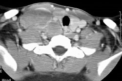
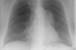
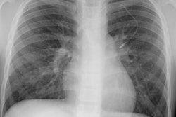
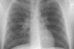
PURPOSE: To determine the appearance of treated thymic lymphoma at magnetic resonance (MR) imaging and to compare the results with findings from posttherapy computed tomography (CT) and clinical outcome. MATERIALS AND METHODS: MR images and CT scans in 25 patients with thymic lymphoma were reviewed. Dimensions of residual masses on MR and CT images were compared. Signal intensity on MR images was assigned to four patterns: (a) low heterogeneous, (b) low homogeneous, (c) high heterogeneous, and (d) high homogeneous. The presence of cysts, calcification, and fatty infiltration was noted. Abnormality of intrathoracic nodes was also documented. RESULTS: Dimensions of residual thymic masses were greater on MR images than on CT scans. The signal intensity pattern was low homogeneous or heterogeneous in 19 patients. Two of these patients had relapse within 1 year. High heterogeneous signal intensity was observed in five patients, four of whom had relapse. Cysts were seen at MR imaging in six patients, of whom one had relapse, but were seen in only one patient at CT. CONCLUSION: Treated thymic lymphoma usually has low signal intensity on MR images regardless of residual mass size, and cysts of high signal intensity may be seen. Relapse occurred mainly in large-volume masses or masses of high heterogeneous signal intensity, indicating that close follow-up or biopsy should be considered in such cases.
PMID: 9114090, MUID: 97268742