J Thorac Imaging 1996;11(3):223-230. Intrathoracic granulocytic sarcomas.
Takasugi JE, Godwin JD, Marglin SI, Petersdorf SH
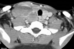
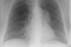
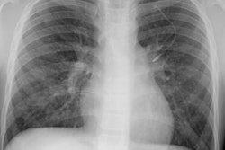
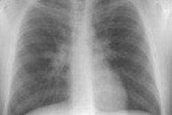
Recent trends in the treatment of intrathoracic granulocytic sarcoma (IGS) call for an overview of its radiographic manifestations. Nine patients from our institution and a review of 41 from the literature provide the basis of our conclusions on the typical and atypical appearance of IGS. Of the nine patients with IGS, all had chest radiographs, five had computed tomographic (CT) scans, and one had magnetic resonance (MR) scans. Radiographic studies and medical records were examined to establish the site and appearance of IGS. Three cases were histologically proved; in the others, the diagnosis was based on clinical presentation and response to chemotherapy. The mediastinum was the most common site of involvement (six of nine cases). A focal mass or mediastinal widening was visible on chest radiographs, and a focal mass or diffuse infiltration or replacement of fat was visible on chest CT. Less common sites of involvement were the lungs (two cases), the pleura (two), the pericardium (two), and the hilar (two). Mediastinal or hilar mass or mediastinal widening is the characteristic finding in IGS. Less common manifestations such as pleural and pericardial effusions and lung opacities should be confirmed histologically, since fluid or tissue is readily accessible.
PMID: 8784735, MUID: 96379172