Nodular Lymphoid Hyperplasia (formerly pseudolymphoma):
Clinical:
Nodular lymphoid hyperplasia represents a well circumscribed
benign
proliferation of lymphoid tissue (a polymorphous population of
both T and B-cells) with numerous reactive germinal centers [1,2].
The diagnosis is controversial and many of the initial cases were
actually MALT
lymphomas [1]. Features which aid in differentiating the lesion
from a MALT
lymphoma include lack of monocytoid B cells and a polyclonal
pattern [1]. Russel bodies- immunoglobulin-containing cytoplasmic
inclusions within plasma cells- may also be present [2].
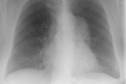
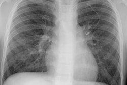
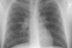
The most common presentation is an incidental finding on CXR (71% of cases) [1]. The lesion is solitary in about two-thirds of cases, but multiple (generally unilateral) in the remaining third [1]. Since it is difficult to completely exclude the diagnosis of a MALT lymphoma, surgical resection is the treatment and is curative [1].
X-ray:
The condition involves only a small area of the lung and appears as a focal subpleural mass or focal mass-like area of consolidation with a mean size of about 2 cm [1,2].
REFERENCES:
(1) Thorax 2001; Travis WD, Galvin JR. Non-neoplastic pulmonary
lymphoid
lesions. 56: 964-71. (No abstract available)
(2) Radiographics 2016; Sirajuddin A, et al. Primary pulmonary lymphoid lesions: radiologic and pathologic findings. 36: 53-70