Photon-counting CT (PCCT) improves the contrast-to-noise ratio of vascular structures imaged on chest CT when compared with conventional CT -- and reduces radiation dose, according to research presented November 26 at the RSNA meeting.
"When compared to conventional … CT scanners, the new technology of [PCCT] will improve quality metrics in contrast-enhanced chest CT by delivering higher contrast-to-noise ratio and at the same time reducing radiation exposure of the patients," said presenter Pooyan Khalighinejad, MD, of the University of Texas Southwestern Medical Center in Dallas.
Khalighinejad's group compared PCCT to energy-integrating detector CT (EID-CT) via a study that included 30 patients referred for routine contrast-enhanced CT, all of whom underwent a PCCT exam and had undergone conventional CT imaging within the past two years (median interval between the two exams, 123 days). The investigators assessed the two techniques' radiation dose (in the lung parenchyma, the thoracic aorta, the main pulmonary artery, the thyroid, the latissimus dorsi, and in mediastinal fat), image noise, and contrast-to-noise ratios. They also collected body mass index (BMI), sex, and age data for each patient and tracked CT dose index (CTDI) and dose length product (DLP).
They reported the following:
| Comparison of conventional CT and PCCT performance for chest imaging | |||
|---|---|---|---|
| Measure | Conventional CT | PCCT | p-value |
| Mean CT dose index volume (CTDIvol) in mGy | 8.4 | 7 | p < 0.01 |
| Dose length product (DLP) in mGy.cm | 299.2 | 258.1 | p < 0.05 |
| Noise (Hounsfield units) | 19.9 | 18.3 | p < 0.05 |
"[PCCT] is an innovative technology that can deliver improved contrast-to-noise ratio of vascular and iodine-rich structures in the chest, while reducing patient radiation exposure," Khalighinejad concluded.
In a related study presented during the same session, a team led by Keiichi Nomura, PhD, of National Cancer Center Hospital East in Kashiwa, Japan, reported that cadmium zinc telluride (CZT)-based PCCT used in super-high-resolution mode improves the diagnostic performance of chest imaging for lung cancer.
"Chest CT scans are routinely performed for diagnosis and staging of lung tumors," Nomura told session attendees. "High-resolution CT images are necessary for more accurate chest diagnosis … [PCCT] has been developed and is under clinical study [for this purpose]."
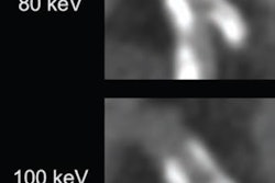
Nomura's group used a prototype PCCT device that included CZT detectors based on an Aquilion Precision system (Canon Medical Systems) with a beam coverage of 40 mm. Patient scans were taken in helical mode at 120 kVp, 250 mA (9.7 mGy), 0.5s rotation, and pitch factor 0.8.
The investigators found that the image noise levels of superhigh-resolution mode and normal-resolution mode were "acceptable for diagnostic tasks," Nomura noted.
"The high-resolution images in the super-high-resolution mode of CZT-[PCCT] contribute to improved diagnostic performance in chest imaging," he concluded.