European healthcare organizations may be facing decisions about new investments in imaging technology for "black swan events" -- that is, battlefield and mass casualty incidents, according to a March 4 discussion at ECR 2026 led by the European Federation of Organizations of Medical Physics.
 Prof. Richard Graham.
Battlefield radiology has long been recognized, but battlefield healthcare in a drone-dominated combat space is quite new, according to U.K. Radiologist Richard Graham, BM BCh, who served in the Royal Naval Reserve and practices at Royal United Hospitals Bath.
"Drones have completely changed warfare," said Graham, who has studied medical imaging infrastructure attacks and the development of battlefield medicine.
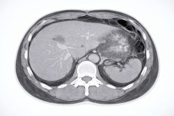
Battlefield medicine distinguishes three roles, according to Graham. Role 1 involves primary care near the point of injury, where point-of-care ultrasound is used most often. Role 2 involves enhanced intermediate care at field clinics, where dynamic digital radiography (DDR) x-ray and ultrasound are key. Role 3 advances care to a specialized hospital model where ultrasound and DDR x-ray are used, but CT is best, he noted.
Mass casualty incidents can quickly overwhelm resources, added Paddy Gilligan, chief physicist at Mater Misericordiae University Hospital in Dublin, Ireland.
 Dr. Paddy Gilligan.
"For the ability of radiology to carry out triage and capacity to add to our healthcare system, we need a plan, and we need not look only within our own hospital, but we need to simulate and do some road testing of that plan for both the acute and chronic phases of the incident," Gilligan advised.
Gilligan has used radiology information system data and architectural designs to develop capacity modeling. Such modeling can also be applied to mass casualty incidents, he said, recommending detailing plans on paper as well, including for the role of the medical physics expert.
Black swan events have occurred around the world, Gilligan explained. When a terror truck was used to attack pedestrians in Nice, France, in 2016, healthcare services conducted 42 CT exams on 42 patients during a two-hour period. In another example, a majority of the tourists involved in a 2016 vehicle collision in Seattle, WA, underwent whole-body CT. Importantly, an earthquake in New Zealand in 2011 led to 21 CTs in six hours -- after a five-hour delay from power outages.
"Power and other infrastructure isn't guaranteed, and that's when we need solutions on the fly," Gilligan said. He listed considerations useful for CT scanner planning in emergency preparedness:
- Table length and weight capacity (tables that can take heavy patients with large, rapid feed)
- Wide bore sizes (>70 cm) and large room sizes (minimum 50 m2)
- High tube heat capacity and dissipation so that tubes do not overheat between patients
- Detector warmup time (can be up to 30 minutes, problematic in emergencies)
- Parallel processing of reconstructions
- Availability of local advanced image processing and storage (imaging networks may not be guaranteed)
- Operating temperatures of scanners (ranges in relation to room cooling requirements)
- Power consumption of CT scanners (simulate and plan in advance)
- Availability of an uninterruptible power supply
Interventional radiology will have the same considerations, but supported by outside teams, the department should plan to supply personal protective equipment, such as lead aprons and glasses.
 Dr. Jonas Andersson, PhD.
Medical Physicist Jonas Andersson, MD, PhD, from Umeå University Hospital in Sweden, highlighted the range of field-based technology, including mobile CT imaging units and C-arms, portable low-field MRI units, and equipment trailers.
Hybrid equipment operating theaters, that enable endovascular resuscitation and trauma management, for example, can save lives, Andersson noted. Regardless, power will be a critical consideration.
Our full coverage of ECR 2026 can be found here.