High-pitch photon-counting CT (PCCT) improves image quality on CT pulmonary angiography (CTPA) for patients with suspected pulmonary embolism (PE) – at lower radiation doses, researchers have found.
The results, published November 15 in the American Journal of Roentgenology, highlight PCCT's potential value for diagnosing suspected PE, according to a team led by Pauline Pannenbecker, MD, of University Hospital Würzburg in Germany.
"CTPA may benefit from [a PCCT] technique," they noted.
Acute PE is a common and often fatal condition, making early detection and treatment crucial, the team noted. CTPA is the go-to test for the diagnostic workup of suspected PE due to benefits such as short acquisition time, availability, and high sensitivity. But dual-energy CTPA with conventional energy-integrating detector technology is limited by its inability to use a high-pitch technique, the group explained.
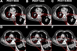
Pannenbecker and colleagues sought to investigate whether high-pitch PCCT CTPA could produce anatomic images and iodine maps of higher quality than conventional CTPA. The team conducted a study that included 117 patients who underwent CTPA for suspected pulmonary embolism (PE) between March 2022 and November 2022. Of these, 58 underwent PCCT CTPA at a pitch of 2, and 59 underwent conventional CTPA at a pitch of 0.55. The investigators reconstructed 120-kV polychromatic images, 60-keV virtual monogenetic images (VMI), and iodine maps for each exam; one radiologist assessed contrast-to-noise and signal-to-noise ratios and three radiologists assessed image quality using a four-point scale (with 1 as the highest quality). The team also tracked radiation dose for each type of CTPA exam.
Pannenbecker and colleagues found that signal-to-noise and contrast-to-noise ratios were higher on PCCT CTPA than conventional CTPA for multicolor images and virtual monochromatic images for all vessels except the left upper lobe artery.
The group also found the following:
| Performance of PCCT CTPA compared with conventional CTPA | |||
|---|---|---|---|
| Measure | Conventional CTPA | PCCT CTPA | p-value |
| Right lower lobe artery, polychromatic images | |||
| Contrast-to-noise ratio | 24.4 | 29.2 | p = 0.001 |
| Signal-to-noise ratio | 28 | 34.5 | p = 0.003 |
| Right lower lobe artery, virtual monochromatic images | |||
| Contrast-to-noise ratio | 29.3 | 37.4 | p = 0.002 |
| Signal-to-noise ratio | 32.7 | 43.2 | p = 0.005 |
Median image quality score for all readers – and for both multicolor and single-color images -- was 1 (i.e., excellent). Image quality of anatomic image sets and iodine maps between the two types of CT exams did not significantly differ, the group reported.
Importantly, PCCT CTPA demonstrated reduced radiation dose, the team noted.
| Radiation dose comparison, conventional CTPA versus PCCT CTPA | |||
|---|---|---|---|
| Measure (median) | Conventional CTPA | PCCT CTPA | p-value |
| CT dose index-volume [mGy] | 4.5 | 3.9 | 0.03 |
| Dose length product [mGy*cm] | 157 | 123.5 | < 0.001 |
| Effective dose [mSv] | 2.8 | 2.2 | < 0.001 |
| Size specific dose estimate [mGy] | 5.3 | 4.6 | 0.01 |
PCCT CTPA's promise as a tool for PE diagnosis merits more research, according to the authors.
"The role of [PCCT] CTPA with reconstruction of iodine maps should continue to be explored for pulmonary embolism detection," they concluded.
The complete study can be found here.