Ultra-high-field 7-tesla (7T) MR spectroscopy has established a significant multivariate relationship between the neurotransmitter glutamate in the brain and sleep patterns, according to a poster presented May 11 at the International Society of Magnetic Resonance Imaging in Medicine (ISMRM) meeting in Cape Town, South Africa.
The study provides insights into the neurochemical basis of sleep disorders in relation to glutamate imbalances and could lead to improved therapeutic management, according to lead author Joshita Majumdar, PhD candidate in electrical and computer engineering, who studied the relationships in a project at the Auburn University Neuroimaging Center in Alabama.
"Glutamate is the brain’s principal excitatory neurotransmitter linked with healthy brain functioning and sleep homeostasis," explained Majumdar for the team led by Gopikrishna Deshpande, PhD, in Auburn's Samuel Ginn College of Engineering. "Deep sleep is associated with a reduction in extracellular glutamate concentrations, reflecting restorative neural processes."
Sleep and neurotransmitters
 Joshita Majumdar, PhD candidate in electrical and computer engineering, presented a snapshot of 7T MRI research based at the Auburn University Neuroimaging Center in Alabama. Other team members not pictured include Nguyen Phuoc Huynh and Adil Bashir, PhD, both of Auburn’s Samuel Ginn College of Engineering, and Lavanya Sankaran of California Health Sciences University in Fresno, CA.Photo courtesy of Joshita Majumdar
Importantly, sleep deprivation has been shown to induce an imbalance in glutamate levels, according to the group. However, sleep-neurotransmitter dynamics have been difficult to determine largely because studies have only examined it through subjective sleep assessments and single-night polysomnography, or experimentally constrained designs, Majumdar noted, adding that studies usually use 3T scanners.
To address these limitations, Majumdar's investigation tracked neurotransmitter levels longitudinally over one year in a single participant. Sleep quality was measured passively using the participant's Oura Ring, a wearable "smart" ring that tracks health metrics.
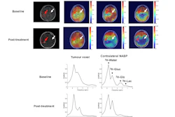
The group acquired proton (1H) MR spectral data using a Magnetom Terra.X 7T MRI scanner (Siemens Healthineers) with a 32-channel head coil. A single-voxel (25 × 20 × 12 mm³) spectrum was obtained from the region of interest, the posterior cingulate cortex (PCC), using a stimulated echo acquisition mode (STEAM) sequence (TR/TE/TM = 3000/20 ms/10 ms, bandwidth = 4 kHz, 128 averages), according to the group.
Although neurotransmitters were not specifically measured during the participant's sleep, a significant correlation observed between prior-night sleep and glutamate concentration over time suggests glutamate-induced sleep regulation, Majumdar explained. The PCC was chosen as the brain region of interest because prior research has linked poor sleep to accelerated volume loss in this area, highlighting its sensitivity to sleep-related metabolic changes.
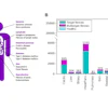
The study is unique in that it followed a naturalistic approach under real-world conditions, without inducing sleep deprivation, said Majumdar, who also highlighted sleep timing, deep sleep duration, and light sleep duration as three key variables. This “deep phenotyping” approach, analyzed with partial least-squares regression, identified a significant relationship (R = 0.31, p < 0.05) between glutamate levels and overall sleep architecture across the 76 weekly scans.
Three variables
"The loading characteristic of the three variables with glutamate indicates that higher glutamate levels correspond to altered sleep, reduced deep sleep, and increased light sleep," Majumdar said. "Together, these findings suggest that increased cortical excitatory activity, reflected by elevated glutamate, is linked to a lighter and less restorative sleep profile."
The findings are consistent with the notion that higher levels of glutamate may impair sleep depth and consolidation, she said.
"Given that sleep is regulated by a multitude of individual-specific factors, the longitudinal design allows us to make stronger inferences about the association," the group concluded.
The project is a snapshot of a larger body of developing work in Alabama, following the installation of a 7T MRI scanner at Auburn University in early 2024. The equipment at Auburn was the first field-installed, clinically approved parallel‑transmit 7T MRI system for patient use, according to the university.
Larger studies are focused on scanning 20 healthy adults twice a week for a full year, while continuously tracking each person’s sleep, physical activity, and nutrition through everyday wearables and apps.
The goal is to understand how the small choices people make every day -- and the changing seasons around them -- shape brain health over time, and to use that knowledge to guide more personalized strategies for preventing and treating conditions such as depression, seasonal affective disorder, and dementia, where disrupted sleep and lifestyle play a central role, according to the group.
Other team members include Nguyen Phuoc Huynh and Adil Bashir, PhD, both of Auburn’s Samuel Ginn College of Engineering, and Lavanya Sankaran of California Health Sciences University in Fresno, CA.
Check out AuntMinnie's full coverage of ISMRM 2026 on our ShowCast.