Researchers have been using quantitative MRI in neurovascular imaging to understand the vasculopathies associated with ischemic stroke.
One example is collaborative research at the University of Washington, University of Alabama at Birmingham (UAB), and University of Utah, where experts are using intracranial vessel wall MRI to differentiate between three intracranial vascular pathologies, explained Niranjan Balu, PhD, co-director of the Vascular Imaging Lab at the University of Washington during a May 14 talk at ISMRM 2026 in Cape Town.
Conventional imaging doesn't very well differentiate intracranial atherosclerosis disease (ICAD), vasculitis, and reversible cerebral vasoconstriction syndrome (RCVS), according to Balu.
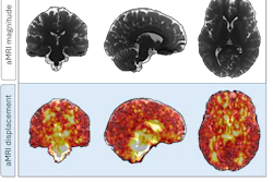
"Qualitative criteria can suffer from inconsistent diagnostic criteria and therefore provide limited reproducibility across studies," he said. Luminal angiograms present as stenosis, Balu explained. On the other hand, high-resolution intracranial vessel wall MR imaging (IVW-MRI) and semiautomated software tools used in combination can differentiate the vasculopathies, as demonstrated in a recent study of 55 patients.
The study evaluated the capability of IVW imaging-derived features to distinguish between ICAD, vasculitis, and RCVS and involved off-label use of gadolinium contrast, Balu noted.
All patients had a clinically confirmed diagnosis of the intracranial vasculopathies -- 25 with intracranial atherosclerosis, 22 with vasculitis, and 8 with RCVS -- and underwent IVW-MRI at a University of Washington hospital on a Prisma 3T scanner (Siemens Healthineers) using a vendor-supplied 64-channel neurovascular coil.
 Niranjan Balu, PhD, and ISMRM
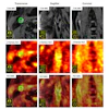
For quantitative image analysis, researchers applied two different types of software: one (VesselVoyager) that draws the center lines of the arteries before they are imported into the second software (MOCHA) that reformats the vessel wall images perpendicular to the artery center lines.
The method identified a total of 279 lesions: 162 atherosclerotic plaques, 94 vasculitis lesions, and 23 RCVS lesions, according to the results. Importantly, the logistic regression model achieved around 79% classification accuracy, with misclassification mainly between atherosclerosis and vasculitis, suggesting quantitative IVW features can facilitate differentiation of diseases, Balu noted.
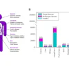
Researchers extracted and calculated the following features: maximum wall thickness, minimum lumen area, eccentricity index (maximum wall thickness–minimum wall thickness/maximum wall thickness), wall volume percentage, and lesion length, he noted.
"Wall volume percentage is used to normalize the wall volume to the sides of the artery and the lesion and basically describe the lesion distribution across the length of the artery," Balu explained.
 Niranjan Balu, PhD, and ISMRM
In terms of quantitative features, ICAD showed the greatest maximum wall thickness (1.95 ± 0.42 mm) and eccentricity (0.64 ± 0.08), compared to vasculitis (1.62 ± 0.36 mm and 0.58 ± 0.08, respectively) and RCVS (1.03 ± 0.23 mm and 0.53 ± 0.07), all p < 0.001 and p < 0.015, respectively, according to the results.
RCVS lesions demonstrated significantly lower percent wall volume compared to atherosclerosis (59.4 ± 7% versus 69.5 ± 9.8%, p < 0.001) and vasculitis (59.4 ± 7% versus 71 ± 6.5%, p < 0.001), while no significant difference was observed between atherosclerosis and vasculitis (p = 0.418), according to the results.
Vasculitis also exhibited significantly higher mean enhancement ratio (1.06 ± 0.34) compared to both atherosclerosis (0.72 ± 0.21, p < 0.001) and RCVS (0.65 ± 0.13, p < 0.001).
The study highlighted that vasculitis showed a more circumferential distribution and highest CE quantitative features, while RCVS looked similar to a normal-looking vessel wall, Balu said.
Key to this research is that it could improve patient management and facilitate objective clinical research, while opening new avenues for evaluation and standardized imaging biomarkers in neurovascular disease, according to the researchers.
"Our findings are also corroborating what we expect from pathophysiology, that atherosclerosis is a focal lesion, vasculitis is primarily inflammatory disease, and RCVS is caused by vasomotor dysfunction with minimal structural change," Balu said.
R01 funding from the U.S. National Institutes of Health (NIH) supported the project. The software used was developed in-house at the University of Washington.
Check out AuntMinnie’s full coverage of ISMRM 2026 on our ShowCast.