Quantitative MRI-based breast density measures are comparable to mammography, according to research presented May 9 at the International Society of Magnetic Resonance in Imaging (ISMRM) annual meeting.
In his presentation, Yasuo Takatsu, PhD, from Fujita Health University in Japan, shared his team’s results showing that quantitative MRI has strong correlation with mammography and superior discrimination in dense breasts.
“It [MRI] offers an objective 3D evaluation overcoming mammography’s limits and provides a foundation for standardized density assessment, personalized screening, and AI-driven risk prediction,” Takatsu said.
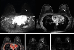
Dense breast tissue creates a masking effect on mammography, potentially hiding cancers on screening for women with intermediate to high breast density. Researchers continue to explore how supplemental imaging such as MRI can help with cancer detection in dense breasts.
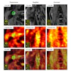
Takatsu and colleagues used 3D MRI to quantitatively visualize breast density. They also compared MRI’s discriminative ability and correlation with mammography. The researchers also studied how MRI-based quantification could overcome mammography’s limitations in this area.
Final analysis for the retrospective study included 107 women with an average age of 61.3 years.
While MRI-derived breast density values were generally lower than those obtained by mammography, Takatsu reported a strong correlation between MRI and mammography (r = 0.83, p < 0.001).
Both modalities showed significant intergroup differences when mammography-based density classification was used as the reference (p < 0.01). Mammography achieved a perfect area under the curve (AUC), but MRI also achieved a high performance (AUC = 0.95, p = 0.24).
However, MRI could significantly differentiate all groups when MRI-based density classification was used as the reference, while mammography could not differentiate fatty from scattered types (p = 0.77).
Additionally, MRI achieved higher performance on receiver operating characteristic (ROC) analysis that considered heterogeneously dense or higher breasts (AUC = 1 versus 0.96, p = 0.04). MRI also achieved a sensitivity of 0.94 and specificity of 0.94 based on BI-RADS density classification. And when MRI classification served as the reference, mammography showed a sensitivity of 0.938 and specificity of 0.852.
Finally, Takatsu said that MRI achieved superior discriminative performance, particularly in the high-density breast group.
Takatsu said the quantitative nature of MRI mitigates interobserver variability that is present in visual BI-RADS assessment.
“Importantly, MRI does not aim to replace mammography in population screening but to complement it," he added. "For women with dense breasts, where mammography sensitivity is known to decrease, MRI-based density quantification could play a key role in risk stratification, guiding individualized screening intervals or supplemental imaging decisions."
Takatsu also said the results could provide foundational data toward integrating MRI-derived metrics into machine-learning pipelines. This could make way for AI-assisted breast density classification and cancer risk prediction.
Check out AuntMinnie’s full coverage of ISMRM 2026 here.