CHICAGO - The chest x-rays alone of a patient suffering from avian flu won't tell a physician that the individual has contracted the deadly disease, according to a radiologist who studied the medical data from 14 Vietnamese victims of the disease.
"Just by looking at the chest x-ray of a person you can't tell that this is caused by influenza A subtype H5N1," said Dr. Nagmi Qureshi, a fellow in thoracic radiology at Churchill Hospital of the University of Oxford in the U.K., in a presentation at the 2005 RSNA meeting. "What you need to know is the person's history: Has he or she been on a farm where there are birds? Have they been in contact with sick birds?"
When you put the x-ray showing multifocal consolidation in the lungs, a representation of pus and infection, and the contact with sick birds together, then, Qureshi said, it is possible to say that the patient may have avian flu.
The investigators studied 98 x-rays of the 14 patients, all of whom were under age 35 and had been admitted to Ho Chi Minh City Hospital in Vietnam. The victims included three children 12 years of age or younger. One of those children survived, she said, but nine of the patients died.
"Characteristically," Qureshi said, "the patients who died came in very sicker, got even sicker and died -- within nine days." The survivors, however, who were hospitalized for as long as 42 days, developed other complications -- pleural effusions and cavitations or areas of deep-seated infections, she said.
 |
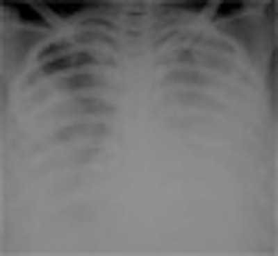
| The H5N1 avian flu virus is endemic in the poultry stocks of nine Southeast Asian countries. Chest x-rays of patients infected with the virus may demonstrate focal consolidation, reticulonodular shadowing, pleural effusion and/or thickening, pneumothorax, lobar collapse, and lymphadenopathy. The appearance and pathological progression of disease on chest x-rays differed between patients who succumbed to avian flu (above) and those who survived infection (below), primarily in terms of continued progression among nonsurvivors, and in the total number of involved segments. Evidence of consolidation in four or more lung segments was significantly associated with mortality. Survivors and nonsurvivors alike were all treated with broad-spectrum antibiotics and the antiviral drug Tamilflu (Roche, Basel, Switzerland). All images courtesy of Dr. Nagmi Qureshi. |
 |
"The x-ray features have some characteristics of both viral and bacterial infection," Qureshi said, "but we cultured no bacteria from any of these patients' sputum or from their blood." The patients were treated with both broad-spectrum antibiotics and antivirals because of the possibility that either type of infection was involved.
"We and others have noted that the pattern of infection among young, healthy individuals -- none of these patients were elderly -- is similar to what has been described as the pattern of the 1918 influenza epidemic, also called the Spanish flu," she said.
Qureshi also compared the avian flu cases with patients who suffered from severe acute respiratory syndrome (SARS). "The appearance of multiple accumulations of infection in the lung is found in both avian flu and SARS," she said. "However, additional abnormalities we discovered in avian flu patients -- including fluid in the space surrounding the lungs, enlarged lymph nodes, and cavities forming in lung tissue -- were absent in patients with SARS."
By Edward Susman
AuntMinnie.com contributing writer
December 2, 2005
Related Reading
X-ray signs correlate with SARS diagnosis and outcome, October 1, 2004
SARS presents distinct radiological patterns, May 6, 2003
Copyright © 2005 AuntMinnie.com