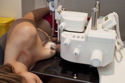
Mammotome has secured clearance from the U.S. Food and Drug Administration (FDA) for two products designed for MR-guided breast biopsy: the Mammotome Prima MR Dual Vacuum-Assisted Breast Biopsy System and the HydroMARK Plus Breast Biopsy Site Marker for MR.
Prima MR is designed to be positioned in the MRI scanner room at the patient's side. It features an 8-gauge needle and touchscreen interface that allows adjustment of aperture size and vacuum strength, according to the company. The system uses 75% less tubing than competing systems, the firm said, enabling larger tissue samples and faster setup and cleanup.
The HydroMARK Plus marker is cleared for exclusive use with the Prima MR system's universal targeting set and incorporates hydrogel technology and titanium shapes that provide visibility under both T1 and T2 MRI sequences, according to Mammotome. The marker is designed to attach to tissue upon deployment and features wings to reduce displacement during surgical excision.
Availability of the products is expected this summer, with international rollout planned for later this year, the company said.

![A normal mammogram confirmed by three-year radiologic follow-up illustrates reader-marked regions of interest (ROIs) during (A) unaided (round 1) and (B) artificial intelligence (AI)–assisted (round 2) reading. Each colored dot represents an ROI for recall by a human reader. Readers could mark more than one ROI per case, represented by multiple dots of the same color. During AI-assisted reading, the AI system displayed three visible prompts: two with suspicion of malignancy scores of 35% (left mediolateral oblique [L MLO] and craniocaudal [L CC]) and one with a suspicion of malignancy score of 10% (right craniocaudal [R CC]), shown as polygonal overlays. Without AI, six of 10 readers (60%) marked a false-positive ROI. With AI assistance, this fell to two of 10 (20%). R MLO = right mediolateral oblique.](https://img.auntminnie.com/mindful/smg/workspaces/default/uploads/2026/07/2026-07-14-radiology-mammogram-ai-auto-bias.H0bYO8QlWs.jpg?auto=format%2Ccompress&fit=crop&h=100&q=70&w=100)


![A normal mammogram confirmed by three-year radiologic follow-up illustrates reader-marked regions of interest (ROIs) during (A) unaided (round 1) and (B) artificial intelligence (AI)–assisted (round 2) reading. Each colored dot represents an ROI for recall by a human reader. Readers could mark more than one ROI per case, represented by multiple dots of the same color. During AI-assisted reading, the AI system displayed three visible prompts: two with suspicion of malignancy scores of 35% (left mediolateral oblique [L MLO] and craniocaudal [L CC]) and one with a suspicion of malignancy score of 10% (right craniocaudal [R CC]), shown as polygonal overlays. Without AI, six of 10 readers (60%) marked a false-positive ROI. With AI assistance, this fell to two of 10 (20%). R MLO = right mediolateral oblique.](https://img.auntminnie.com/mindful/smg/workspaces/default/uploads/2026/07/2026-07-14-radiology-mammogram-ai-auto-bias.H0bYO8QlWs.jpg?auto=format%2Ccompress&fit=crop&h=112&q=70&w=112)