The requirements for successful advancements in 3D software vary from application to application. There are generic requirements in the areas of interactivity, usability, segmentation, and registration capabilities of a system. Particular applications also have more specific requirements that demand justification for the extra effort and expense involved in refining a system's usability. To a large extent, these specific requirements depend on the medical purpose for which the software is being used.
Interactivity
Traditionally, the 3D display of medical datasets has been hampered by the computation-intensive and time-consuming processes necessary to create such images. Advances today in high-end computer hardware and graphic board designs have overcome most such limitations.
Interactivity in 3D is important because the physician has no way of predicting the desirable angle for viewing pathological abnormalities. For each case, a large number of thresholding or mapping functions must be implemented to gather information from the 3D image. In some applications, such as intraoperative navigation, telesurgery, and surgical training, real-time feedback at 30 frames per second (fps) is crucial. In most other cases, for the physician to be able to manipulate the data as quickly and simply as possible, the system must at least be interactive (5 fps).
Accurate models
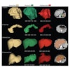
One of the biggest challenges of 3D reconstruction is guaranteeing that the volumetric model resembles the actual anatomical structures. Unfortunately, at this time no set standards exist for segmentation techniques or the parameters for extracting a region of interest from imaging modalities. Using different segmentation parameters can result in differently reconstructed models. Even in the case of a simple window leveling, a slight change in threshold values can result in significant changes in the final structures.
The lack of accuracy in 3D models for applications such as diagnosis, postoperative validation, and patient screening should be treated very seriously because they could result in misdiagnosis, posing liability issues. For such applications, the segmentation process must be very specific to the pathology. Such segmentations are usually conducted semiautomatically by the physician on a 2D or 3D image prior to reconstruction.
For applications such as preoperative planning, intraoperative navigation, or telesurgery, in which the pathology has been diagnosed prior to 3D reconstruction, the segmentation issue is not as crucial because the objective is not diagnostics. However, even in these cases, segmentation is still very important. The main objective in such applications is to look at the spatial relationships between the 3D structures for the purpose of functional restoration and assessment of the best surgical path to the target lesion.
In teaching and training applications, the 3D model should resemble the characteristics of the actual anatomical model in its elasticity, stiffness, and interactions with other organs. One way to produce accurate 3D models in the future is to introduce segmentation and reconstruction standards, which would be specific to the region of interest on a given modality.
Correlation
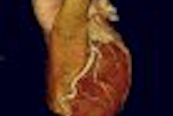
Correlating signals obtained by various modalities could be one of the most important features of 3D software. A robust 3D viewing application should be able to superimpose or register volumetric signals from different imaging sources. This is necessary for tasks such as comparing various signals obtained from different modalities and observing the time evolution of a given pathology.
The main applications of volume-with-volume registration are for segmentation, diagnostics, and screening. Accurate and reproductive methods of registration independent of patient positioning are required to perform context fusion and scaling.
Patient-to-image registration is the basis for localizing the pathology within the body (stereotactic surgery). This type of registration is fairly accurate and easy to implement because it utilizes a frame of reference or reference markers, which are attached to the patient's head, from the time of imaging to the operation. The main applications of stereotaxi are intraoperative planning, intraoperative navigation, and telesurgery.
User interface
Finally, the user interface is probably the most crucial part of deploying clinical software since only it can determine whether the software will be utilized effectively. In designing a clinical user interface, one has to bear in mind that the software must conform to the physician's needs, not vice versa. When using the 3D model, the physician should be concerned with the task of identifying the clinical problem or selecting which surgical procedure to apply, rather than how to use the software.
It is crucial that the user interface not burden the physician with the details of implementing the computational model. All aspects of graphics and analysis should be carried out automatically. To maintain simplicity without losing functionality, the user interfaces must be specifically designed for a given task. Every diagnostic problem requires its own analysis tools and each with its own interface. For instance, cardiac requires different tools than orthopedics. If one tries to design a single generic universal diagnostic software, chances are that it will either become very complex or it will lack significant functionality.
If the industry addresses these issues in a cooperative, multidisciplinary manner, rapid advances are possible. This cooperation must extend to modality and software vendors, research institutions, and physicians from around the globe from all relevant specialties. Getting broad cooperation is always difficult, but if it is achieved the practice of medicine can continue to have multidimensional advances.
By Ramin Shahidi, Ph.D.
AuntMinnie.com contributing writer
November 8, 2006
Reprinted from Maximum Vision magazine by permission of Emageon. Ramin Shahidi, Ph.D., is the director of Image Guidance Laboratories (IGL) and a faculty in the department of surgery at Stanford University in Stanford, CA. Shahidi has authored more than 50 scientific publications in the field of medical imaging and currently serves as a deputy editor for the Journal of Computer Assisted Radiology and Surgery (JCARS) and is on the editorial board of the Journal of Computer Aided Surgery (JCAS).
Related Reading
Part II: Medical image processing has room to grow, September 4, 2006
3D penetrates trauma imaging niche, August 24, 2006
3D PET/CT demonstrates virtual vigor, July 27, 2006
Part I: Medical image processing has room to grow, July 4, 2006
Cardiac SPECT/CT fusion captures SNM's Image of the Year, June 6, 2006
Copyright © 2006 Emageon