Morphological markers culled from diffusion tensor MRI (DT-MRI) brain scans using an AI algorithm can help diagnose autism in young children with an accuracy rate of 98.5%, according to research to be presented at the upcoming RSNA meeting.
The findings could translate to earlier diagnosis of the condition in young children, according to study co-author Gregory Barnes, MD, PhD, of the Norton Children's Autism Center in Louisville, Kentucky.
"Imaging offers the promise of quickly detecting autism in an objective fashion," Barnes said in a statement released November 21 by the RSNA. "We envision an autism assessment that begins with DT-MRI followed by an abbreviated session with a psychologist to confirm the results and guide parents on next steps. This approach could reduce the psychologists' workload by up to 30%."
Fewer than half of children with autism spectrum disorder undergo a developmental evaluation by three years of age, and 30% of children who met the criteria for autism spectrum disorder do not receive a formal diagnosis by eight years of age, the team noted, citing the Centers for Disease Control and Prevention's (CDC) 2023 Community Report on Autism. Identifying autism early allows for better treatment.
"The idea behind early intervention is to take advantage of brain plasticity, or the ability of the brain to normalize function with therapy," Barnes said.
Lead author Mohamed Khudri of the University of Louisville and colleagues investigated whether morphological markers extracted from DT-MRI scans had any correlation with Social Responsiveness Scale (SRS) behavioral scores and thus could classify individuals as having autism spectrum disorder or as being "typically developing" individuals. The team conducted a study that included data from the Autism Brain Imaging Data Exchange-II for 226 children between the ages of two and three. Of these 226 children, 126 had autism and 100 did not.
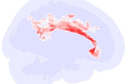
 The top five white matter features (region pairs) in a single image. The color map is: yellow = superior cerebellar peduncle (R)/uncinate fasciculus (R), orange = column and body of fornix/posterior corona radiata (L), purple = splenium/retrolenticular internal capsule (L), blue = dorsal cingulum (L)/cres of fornix (R), green = splenium/external capsule (R). Image and caption courtesy of the RSNA.
An AI software algorithm collected brain tissue images from the MRI scans and extracted markers that indicated connectivity between brain regions, while a deep-learning algorithm compared the marker patterns between children with autism and those without the condition.
The AI/deep-learning technology demonstrated 97% sensitivity, 98% specificity, and an overall accuracy of 98.5% for identifying the children with autism.
"Our approach is a novel advancement that enables the early detection of autism in infants under two years of age," Khudri said in the RSNA statement. "We believe that therapeutic intervention before the age of three can lead to better outcomes, including the potential for individuals with autism to achieve greater independence and higher IQs."
The researchers are working toward securing clearance from the U.S. Food and Drug Administration for the software, they said.