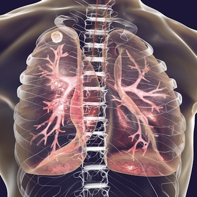
A deep learning-based tuberculosis (TB) detection model can detect TB on phone-captured chest x-ray photographs, a poster presented at the RSNA 2020 virtual conference revealed.
A research team led by Po-Chih Kuo, PhD, an assistant professor of computer science from National Tsing Hua University in Taiwan, explained how the algorithm TBShoNet can be used on phones to help diagnose TB in areas where radiologists and high-resolution digital images are unavailable.
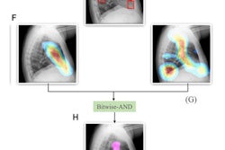
The neural network was pretrained on a database of 250,044 chest x-rays with 14 pulmonary labels that did not include TB. TBShoNet was then recalibrated for the use of chest x-ray photographs by using simulation methods. Model performance was tested using 662 chest x-ray photographs taken by five different phones (336 TB cases and 326 normal chest x-rays).
The researchers found TBShoNet produced 81% sensitivity and 84% specificity.





![A normal mammogram confirmed by three-year radiologic follow-up illustrates reader-marked regions of interest (ROIs) during (A) unaided (round 1) and (B) artificial intelligence (AI)–assisted (round 2) reading. Each colored dot represents an ROI for recall by a human reader. Readers could mark more than one ROI per case, represented by multiple dots of the same color. During AI-assisted reading, the AI system displayed three visible prompts: two with suspicion of malignancy scores of 35% (left mediolateral oblique [L MLO] and craniocaudal [L CC]) and one with a suspicion of malignancy score of 10% (right craniocaudal [R CC]), shown as polygonal overlays. Without AI, six of 10 readers (60%) marked a false-positive ROI. With AI assistance, this fell to two of 10 (20%). R MLO = right mediolateral oblique.](https://img.auntminnie.com/mindful/smg/workspaces/default/uploads/2026/07/2026-07-14-radiology-mammogram-ai-auto-bias.H0bYO8QlWs.jpg?auto=format%2Ccompress&fit=crop&h=112&q=70&w=112)


