Dear AuntMinnie Member,
People who contracted then recovered from COVID-19 often continue to experience symptoms, even months after the initial infection. Clinicians are trying to track these long-term effects, and MRI offers a good way to do that.
In our top-viewed story this week, researchers describe how susceptibility-weighted MRI can illuminate brain changes in people who have recovered from COVID-19 up to six months later, including symptoms such as difficulty concentrating, headache, sleep disruptions, and depression or anxiety.
In addition to this top story, research that will be presented at the upcoming RSNA meeting demonstrates how ultrahigh-resolution MRI shows enlarged perivascular spaces in the brains of people who suffer from migraines -- and how an MRI neuroimaging-based machine-learning model could help clinicians diagnose attention deficit hyperactivity disorder.
You can access these articles as well as other coverage of the modality in our MRI Community.
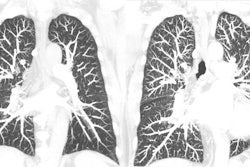
Low-dose CT for lung cancer screening
It's been shown that annual low-dose CT for lung cancer screening reduces mortality rates, and our second most-viewed story of the week adds further evidence to this, finding an 80% overall 20-year survival rate for individuals diagnosed with lung cancer when the disease is caught early.
Also take a look at our coverage of how Ukrainian radiologists are coping with providing care in the midst of war, dealing with the significant challenge of stabilizing patients quickly, imaging them, and then transferring them to safer care centers.
Check out our CT Community to find out more.
The Road and the Radcast
RSNA 2022 is upon us! Meeting attendees will gather in Chicago on Sunday, November 27, to renew professional relationships -- and add new ones -- take in a raft of scientific presentations, and enjoy the frosty, almost-winter beauty of the city. Check out our preview coverage of the conference: Artificial Intelligence, Women's Imaging, Digital X-ray, MRI, Ultrasound, Molecular Imaging, and CT. (Also, keep your eyes peeled for Imaging Informatics, which will post on November 25.)
Once the meeting starts, AuntMinnie.com will be covering it live with our RadCast @ RSNA 2022. Check out the site regularly throughout the week -- you won't want to miss it.