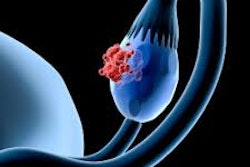
Deep learning (DL) can help improve ovarian lesion classification from MRI exams, according to research published August 5 in Radiology.
Investigators led by Wen-Chi Hsu, MD, from Johns Hopkins University in Baltimore, MD, found that their DL approach accurately differentiated malignant from benign ovarian lesions on MRI scans. It also reduced segmentation time and classified lesions on par with radiologists.
"Clinically, [the model] could serve as a second-reader system, assisting radiologists with segmentation and classification outputs to cross-check their assessments," Hsu and colleagues wrote.
MRI can characterize complex masses by offering tissue contrast that is superior to ultrasound. And while previous studies have shown promise for MRI-based DL models, the researchers noted that few externally validated models are available.
Hsu and colleagues developed a comprehensive end-to-end pipeline for MRI-based classification of ovarian lesions. They integrated Meta’s Segment Anything Model (SAM) for automatic lesion segmentation with a DenseNet-121 DL model incorporating both imaging and clinical data. The researchers then trained and validated the model for ovarian lesion classification.
 (A) Images depict the workflow of the Segment Anything Model (SAM). In a 29-year-old woman with a right ovarian mass subsequently confirmed as well-differentiated mucinous carcinoma, a bounding box (red) is placed around the lesion on axial T1-weighted contrast-enhanced (T1CE) and axial T2-weighted (T2WI) MRI scans, allowing the SAM to automatically generate a segmentation mask (green). (B) Flowchart shows the integration of preprocessed T1-weighted contrast-enhanced (T1CE) and T2-weighted (T2WI) segmented images alongside clinical data into the deep-learning (DL) model for lesion classification. (C) Flowchart shows dual-path network architecture for ovarian lesion classification, featuring components such as a multilayer perceptron (MLP), three-dimensional convolutional neural network (3D CNN), dynamic nonlinear (D-NonL) modules, elementwise addition (+), upsampling (U), and concatenation (C).RSNA
Radiologists, meanwhile, classified lesions based on O-RADS, based on subjective assessment. The team trained the model using datasets from three institutions, including two from the U.S. and one from Taiwan.
The primary dataset included 534 lesions from 448 women with a mean age of 52 from one U.S. institution. External datasets included 58 lesions from 55 women with a mean age of 51 from another U.S. institution and 29 lesions from 29 women with an average age of 49 from an institution in Taiwan.
The team reported the following findings:
Segmentation by SAM achieved a Dice coefficient of 0.86 to 0.88.
SAM-assisted segmentation reduced the processing time per lesion by four minutes compared with manual segmentation.
The DL classification model achieved an area under the receiver operating characteristic curve (AUC) of 0.85 on the internal test and 0.79 across both external datasets with SAM-segmented images. Radiologists achieved an AUC of 0.84 (p > 0.05).
The segmentation accuracy results of the SAM model underscores could potentially balance automation and precision in MRI-based ovarian lesion segmentation, the study authors highlighted.
"These findings support integrating the SAM for segmentation with DL models for classification, offering a promising end-to-end pipeline with strong potential for clinical translation," they wrote.
The results represent a "compelling demonstration" of how foundation models can improve radiology workflows, according to an accompanying editorial written by Rajesh Bhayana, MD, from the University of Toronto, and Bo Wang, PhD, from Toronto General Hospital Research Institute.
The two highlighted that while full autonomy has yet to be reached, the results reflect a shift toward modular AI systems that can be deployed flexibly and upgraded regularly with radiologists involved.
"The broader implication is that foundation models and radiologists will increasingly work together, in tandem, to streamline imaging interpretation and patient management," Bhayana and Wang wrote.
The full study can be found here.