
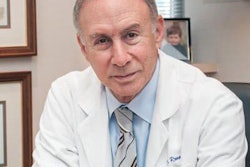
The University of California, San Francisco (UCSF) has tapped Dr. Christopher Hess, PhD, to lead its department of radiology and biomedical imaging, replacing Dr. Ronald Arenson, who retired in October.
Dr. Christopher Hess, PhD.Hess is currently a professor of radiology and neurology and chief of neuroradiology at UCSF. He also serves as the associate chair for quality and safety at the department, and he is associate director of the U.S. National Institutes of Health's T32 training grant program.
Hess joined the faculty in 2008 after completing a radiology residency and neuroradiology fellowship at UCSF. He earned his medical degree at the University of Illinois, where he also received bachelor's and master's degrees and a doctorate in electrical engineering.
Hess holds leadership positions at the RSNA, the International Society for Magnetic Resonance in Medicine (ISMRM), and the American Society of Neuroradiology. He also serves on the editorial board of the American Journal of Neuroradiology and is incoming deputy editor for Radiology.
His areas of research include MRI of brain degeneration, development, and vascular disease. He has also served as fellowship director of neuroradiology at UCSF, and he teaches in the university's Undergraduate Medical Education and Masters of Science in Biomedical Imaging programs, as well as in postgraduate education programs for radiology and neurosurgery.


![Overview of the study design. (A) The fully automated deep learning framework was developed to estimate body composition (BC) (defined as subcutaneous adipose tissue [SAT] in liters; visceral adipose tissue [VAT] in liters; skeletal muscle [SM] in liters; SM fat fraction [SMFF] as a percentage; and intramuscular adipose tissue [IMAT] in deciliters) from MRI. The fully automated framework comprised one model (model 1) to quantify different BC measures (SAT, VAT, SM, SMFF, and IMAT) as three-dimensional (3D) measures from whole-body MRI scans. The second model (model 2) was trained to identify standardized anatomic landmarks along the craniocaudal body axis (z coordinate field), which allowed for subdividing the whole-body measures into different subregions typically examined on clinical routine MRI scans (chest, abdomen, and pelvis). (B) BC was quantified from whole-body MRI in over 66,000 individuals from two large population-based cohort studies, the UK Biobank (UKB) (36,317 individuals) and the German National Cohort (NAKO) (30,291 individuals). Bar graphs show age distribution by sex and cohort. BMI = body mass index. (C) After the performance assessment of the fully automated framework, the change in BC measures, distributions, and profiles across age decades were investigated. Age-, sex-, and height-adjusted body composition reference curves were calculated and made publicly available in a web-based z-score calculator (https://circ-ml.github.io).](https://img.auntminnie.com/mindful/smg/workspaces/default/uploads/2026/05/body-comp.XgAjTfPj1W.jpg?auto=format%2Ccompress&fit=crop&h=112&q=70&w=112)


