Dear AuntMinnie Member,
CHICAGO - The RSNA show opened today, with the cozy confines of McCormick Place keeping conference attendees dry from the rainy weather outside. And in what's become an annual tradition, your AuntMinnie editorial staff is on hand to provide daily updates on the latest clinical and business news in radiology with our RADCast@RSNA special section.
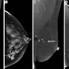
Breaking news we're featuring today include a new study on the use of MR spectroscopy for breast imaging. U.S. researchers found that breast MRS caught all cases of invasive carcinoma in their study population, and was not prone to false-positive readings. Read all about it by clicking here.
In another story, Canadian researchers examined the utility of contrast-enhanced ultrasound to differentiate metastases from primary liver tumors by examining factors like time to peak enhancement and washout. Learn more by clicking here.
Also, U.S. researchers reported on the results of the American College of Radiology Imaging Network/Gynecology Oncology Group (ACRIN/GOG) A6651 study, which examined the use of MRI for diagnosing invasive cervical cancer. They found it as useful as CT -- find out why by clicking here.Keep checking back with the RADCast, at radcast.auntminnie.com, through RSNA week for more late-breaking reports from medical imaging's showcase meeting. And while you're visiting, try out our new Question of the Week poll, on a topic that everyone seems to have an opinion on -- the timing of the RSNA show itself. Should it stay in Chicago, or relocate to sunnier climes? Let us know by going to question.auntminnie.com.
![Images show the pectoralis muscles of a healthy male individual who never smoked (age, 66 years; height, 178 cm; body mass index [BMI, calculated as weight in kilograms divided by height in meters squared], 28.4; number of cigarette pack-years, 0; forced expiratory volume in 1 second [FEV1], 97.6% predicted; FEV1: forced vital capacity [FVC] ratio, 0.71; pectoralis muscle area [PMA], 59.4 cm2; pectoralis muscle volume [PMV], 764 cm3) and a male individual with a smoking history and chronic obstructive pulmonary disorder (COPD) (age, 66 years; height, 178 cm; BMI, 27.5; number of cigarette pack-years, 43.2, FEV1, 48% predicted; FEV1:FVC, 0.56; PMA, 35 cm2; PMV, 480.8 cm3) from the Canadian Cohort Obstructive Lung Disease (i.e., CanCOLD) study. The CT image is shown in the axial plane. The PMV is automatically extracted using the developed deep learning model and overlayed onto the lungs for visual clarity.](https://img.auntminnie.com/mindful/smg/workspaces/default/uploads/2026/03/genkin.25LqljVF0y.jpg?auto=format%2Ccompress&crop=focalpoint&fit=crop&h=100&q=70&w=100)


![Images show the pectoralis muscles of a healthy male individual who never smoked (age, 66 years; height, 178 cm; body mass index [BMI, calculated as weight in kilograms divided by height in meters squared], 28.4; number of cigarette pack-years, 0; forced expiratory volume in 1 second [FEV1], 97.6% predicted; FEV1: forced vital capacity [FVC] ratio, 0.71; pectoralis muscle area [PMA], 59.4 cm2; pectoralis muscle volume [PMV], 764 cm3) and a male individual with a smoking history and chronic obstructive pulmonary disorder (COPD) (age, 66 years; height, 178 cm; BMI, 27.5; number of cigarette pack-years, 43.2, FEV1, 48% predicted; FEV1:FVC, 0.56; PMA, 35 cm2; PMV, 480.8 cm3) from the Canadian Cohort Obstructive Lung Disease (i.e., CanCOLD) study. The CT image is shown in the axial plane. The PMV is automatically extracted using the developed deep learning model and overlayed onto the lungs for visual clarity.](https://img.auntminnie.com/mindful/smg/workspaces/default/uploads/2026/03/genkin.25LqljVF0y.jpg?auto=format%2Ccompress&crop=focalpoint&fit=crop&h=112&q=70&w=112)


