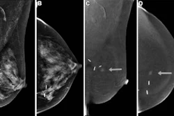
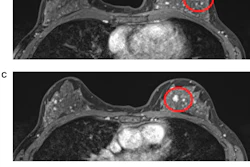
Contrast-enhanced mammography (CEM) has been on the rise in recent years, with recent studies showing that it may be a suitable substitute for breast MRI in some women.
Wendie Berg, MD, PhD, has been studying CEM's utility, including for women with dense breasts and those with a personal history of breast cancer. Berg is a professor of radiology at the University of Pittsburgh School of Medicine and a researcher at the Hillman Cancer Center at the University of Pittsburgh Medical Center.
Berg presented research at RSNA 2025 showing that adding digital breast tomosynthesis (DBT) to CEM leads to some decreases in false-positive recalls.
Wendie Berg, MD, PhD, discusses current barriers surrounding the use of CEM.
She also co-founded DenseBreast-info.org, an educational resource for clinicians and patients on how breast density affects women’s breast cancer risk.
Berg talks about the advantages and considerations for CEM, how the modality ties into the work being done at DenseBreast-info.org, and results from recent CEM studies.
View the full episode below.
![A normal mammogram confirmed by three-year radiologic follow-up illustrates reader-marked regions of interest (ROIs) during (A) unaided (round 1) and (B) artificial intelligence (AI)–assisted (round 2) reading. Each colored dot represents an ROI for recall by a human reader. Readers could mark more than one ROI per case, represented by multiple dots of the same color. During AI-assisted reading, the AI system displayed three visible prompts: two with suspicion of malignancy scores of 35% (left mediolateral oblique [L MLO] and craniocaudal [L CC]) and one with a suspicion of malignancy score of 10% (right craniocaudal [R CC]), shown as polygonal overlays. Without AI, six of 10 readers (60%) marked a false-positive ROI. With AI assistance, this fell to two of 10 (20%). R MLO = right mediolateral oblique.](https://img.auntminnie.com/mindful/smg/workspaces/default/uploads/2026/07/2026-07-14-radiology-mammogram-ai-auto-bias.H0bYO8QlWs.jpg?auto=format%2Ccompress&fit=crop&h=100&q=70&w=100)


![A normal mammogram confirmed by three-year radiologic follow-up illustrates reader-marked regions of interest (ROIs) during (A) unaided (round 1) and (B) artificial intelligence (AI)–assisted (round 2) reading. Each colored dot represents an ROI for recall by a human reader. Readers could mark more than one ROI per case, represented by multiple dots of the same color. During AI-assisted reading, the AI system displayed three visible prompts: two with suspicion of malignancy scores of 35% (left mediolateral oblique [L MLO] and craniocaudal [L CC]) and one with a suspicion of malignancy score of 10% (right craniocaudal [R CC]), shown as polygonal overlays. Without AI, six of 10 readers (60%) marked a false-positive ROI. With AI assistance, this fell to two of 10 (20%). R MLO = right mediolateral oblique.](https://img.auntminnie.com/mindful/smg/workspaces/default/uploads/2026/07/2026-07-14-radiology-mammogram-ai-auto-bias.H0bYO8QlWs.jpg?auto=format%2Ccompress&fit=crop&h=112&q=70&w=112)