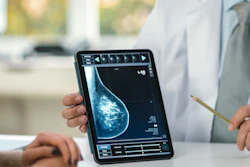
An AI-assisted computer-aided diagnosis (CAD) system significantly improved the diagnostic performance of breast radiologists reading mammograms, with specificity rising from 77% to 88.4%, researchers have reported.
The study also found that average interpretation time fell by more than 30%, noted a team led by Wu Lin Low Ong, MD, of the National Cancer Center in Singapore. The findings were published April 22 in Academic Radiology.
"AI-CAD significantly enhances specificity and reduces reading time in mammographic interpretation without compromising sensitivity," the group wrote.
The research was prompted by the fact that the Asia-Pacific region bears a disproportionate breast cancer burden. The authors noted that, in 2022, the age-standardized incidence rate reached 34.3 per 100,000 against an age-standardized mortality rate of 10.5 per 100,000 -- a ratio that reflects late-stage diagnosis patterns linked to limited screening access.
"Many Asian countries experience high incidence-to-mortality ratio due to limited organized screening programs, resource constraints and manpower limitations," Ong and colleagues wrote. "The application of AI in mammography interpretation may help address these challenges."
The group conducted a study that included nine experienced breast radiologists from institutions across Singapore, the Philippines, India, Egypt, and Hong Kong. Each reader interpreted the same set of 302 digital mammograms -- which included 89 biopsy-proven breast cancers -- in two separate sessions, one unassisted and one with AI-CAD support.
The team reported the following:
- With AI assistance, the average specificity increased from 77% to 88.4% (p = 0.03).
- The average area under the receiver operating characteristic curve (AUROC) for accuracy rose from 0.799 without use of AI-CAD to 0.851 with its use (p = 0.0151).
- Sensitivity did not change significantly between sessions, suggesting that the gains in specificity were not achieved at the cost of cancer detection.
- Workflow efficiency improved substantially, with mean interpretation time per case dropping 32%, from 121.5 seconds without assistance from AI-CAD to 83.2 seconds with it (p < 0.001).
Ong and colleagues concluded that AI-CAD can enhance diagnostic efficiency in mammographic interpretation without compromising sensitivity and support its integration into breast cancer screening workflows. They did underscore, however, "the importance of maintaining human oversight and critical judgment when using AI in clinical practice."
Access the full study here.