A Swedish study of nearly 15,000 women found that digital breast tomosynthesis (DBT) had a 1.6% false-positive recall rate, higher than digital mammography but lower than previous DBT trials, according to research published in European Radiology.
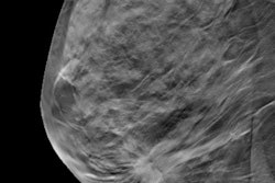
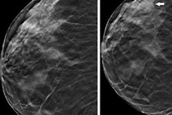
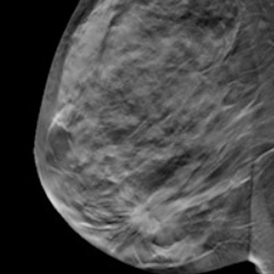
A team led by Dr. Kristin Johnson from Lund University in Malmö concluded in the Malmö Breast Tomosynthesis Screening Trial (MBTST) that the higher false-positive rate was mainly caused by an increased detection of stellate findings on DBT, which include distortions and a density with stellate (star-shaped) configuration. However, this could also point to a learning curve for radiologists using DBT.
"The proportion of these [stellate] findings was reduced after the first trial year," Johnson and colleagues wrote.
While research continues to show DBT's promise in breast cancer screening, the study authors cautioned that false-positive cases from the modality should be studied to better understand potential consequences. They also noted that prospective European trials have shown both higher and lower false-positive recall rates in screening with DBT compared with digital mammography. However, retrospective trials from the U.S. have shown lower false-positive recall rates compared with mammography.
MBTST is a prospective, population-based trial with 14,848 participating women. It was designed to compare one-view DBT to two-view digital mammography in breast cancer screening.
Using data from the trial, Johnson and co-authors wanted to evaluate the total number of false-positive recalls on DBT, including radiographic appearances and false-positive biopsies, as well as compare findings to that of digital mammography.
The researchers found that DBT had a false-positive recall rate of 1.6% compared with 0.8% seen with screening digital mammography. They also reported that the false-positive rate in the DBT was at its highest the first year of the trial at 2.6%. After that, it stabilized at around 1.5%. However, the false-positive rate in the mammography group varied between 0.5% and 1% throughout the trial.
The team also found that the proportion of the radiographic appearance of stellate distortion was 37.3% with DBT and 24% with mammography. For the first year of the trial, when the DBT false-positive rate was at its highest, the researchers found that the percentage of stellate distortion with DBT was 50% (19/38) compared to 35% (72/206) for the remaining trial years.
The study authors suggested that the decrease in proportion of stellate distortions over time could mean a learning curve in interpreting these imaging findings.
"Increased experience, in combination with access to prior DBT examinations, could decrease the false-positive recall rates in the future," they wrote.
Johnson and colleagues also called for more studies on false-positive recalls and false-positive appearances in subsequent screening rounds. They wrote that these could help breast radiologists learn how access to prior DBT can affect false positives in such screening.