The American College of Radiology (ACR) is teaming up with GE Healthcare and the Breast Cancer Research Foundation to launch a clinical trial to determine whether contrast-enhanced mammography (CEM) improves breast cancer detection for women with dense breasts.
The Contrast-Enhanced Mammography Imaging Screening Trial (CMIST) will seek to determine if CEM provides more accurate cancer detection compared with digital breast tomosynthesis (DBT), as well as reduce false-positive exams in women with dense breasts, according to the ACR.
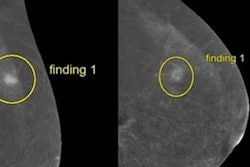
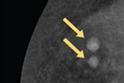
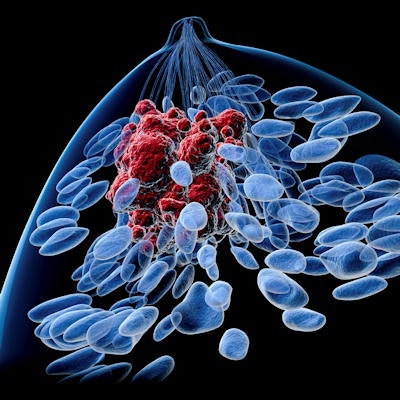
CEM combines mammography and vascular-based screening methods to highlight areas of unusual blood flow patterns that may indicate malignancy. Early studies of CEM in screening women with dense breasts have shown the potential benefit of CEM in the detection of breast cancer, the ACR noted.
The planned study will be managed by ACR's Center for Research and Innovation, with support from the Breast Cancer Research Foundation and GE. Dr. Christopher Comstock of Memorial Sloan Kettering Cancer Center will serve as the principal investigator.

![A normal mammogram confirmed by three-year radiologic follow-up illustrates reader-marked regions of interest (ROIs) during (A) unaided (round 1) and (B) artificial intelligence (AI)–assisted (round 2) reading. Each colored dot represents an ROI for recall by a human reader. Readers could mark more than one ROI per case, represented by multiple dots of the same color. During AI-assisted reading, the AI system displayed three visible prompts: two with suspicion of malignancy scores of 35% (left mediolateral oblique [L MLO] and craniocaudal [L CC]) and one with a suspicion of malignancy score of 10% (right craniocaudal [R CC]), shown as polygonal overlays. Without AI, six of 10 readers (60%) marked a false-positive ROI. With AI assistance, this fell to two of 10 (20%). R MLO = right mediolateral oblique.](https://img.auntminnie.com/mindful/smg/workspaces/default/uploads/2026/07/2026-07-14-radiology-mammogram-ai-auto-bias.H0bYO8QlWs.jpg?auto=format%2Ccompress&fit=crop&h=100&q=70&w=100)


![A normal mammogram confirmed by three-year radiologic follow-up illustrates reader-marked regions of interest (ROIs) during (A) unaided (round 1) and (B) artificial intelligence (AI)–assisted (round 2) reading. Each colored dot represents an ROI for recall by a human reader. Readers could mark more than one ROI per case, represented by multiple dots of the same color. During AI-assisted reading, the AI system displayed three visible prompts: two with suspicion of malignancy scores of 35% (left mediolateral oblique [L MLO] and craniocaudal [L CC]) and one with a suspicion of malignancy score of 10% (right craniocaudal [R CC]), shown as polygonal overlays. Without AI, six of 10 readers (60%) marked a false-positive ROI. With AI assistance, this fell to two of 10 (20%). R MLO = right mediolateral oblique.](https://img.auntminnie.com/mindful/smg/workspaces/default/uploads/2026/07/2026-07-14-radiology-mammogram-ai-auto-bias.H0bYO8QlWs.jpg?auto=format%2Ccompress&fit=crop&h=112&q=70&w=112)