Tuesday, November 30 | 8:00 a.m.-9:00 a.m. | SSBR05-5 | Room TBA
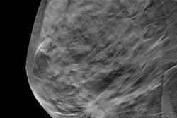
In this presentation, Dr. Stamatia Destounis from Elizabeth Wende Breast Care will talk about the effects of adding mammographic density to the Tyrer-Cuzick risk model.Destounis will present findings from her team's study of over 59,000 women who underwent full-risk assessment between July 2019 and January 2020 using version 7 of the model at the time of screening at a specialized breast imaging clinic.
The researchers found that compared with the 8.3% of women classified as high risk using the model with no information input on breast density, there were significantly higher proportions of high-risk women when using their own "version 8" of the model, which adds volumetric breast density calculations to the mix.
Younger, premenopausal women were found to be more affected by this addition via higher odds ratios. The study authors also found that over one-third of low- or intermediate-risk women have dense breasts and may still benefit from supplemental imaging.
Learn what else the team found, as well as how clinics can use this information, at this morning session.