
Tuesday, November 30 | 9:30 a.m.-10:30 a.m. | SSPH09-5 | Room TBA
In this Tuesday morning presentation, research findings will show how high-resolution photon-counting CT used with deep-learning image processing can expand the modality's role in breast cancer imaging.CT isn't typically used for breast cancer imaging due to spatial and contrast resolution limitations, noted a team led by doctoral candidate Nathan Huber of Mayo Clinic Graduate School of Biomedical Sciences in St. Peter, MN. But new technology such as high-resolution, whole-body photon-counting CT -- combined with deep learning to process the images -- shows promise for visualizing microcalcifications.
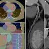
The group used a work-in-progress whole-body photon-counting CT scanner from Siemens Healthineers to image a patient with a history of breast cancer. These images were then processed by the deep-learning algorithm and compared with the patient's prior mammography exam.
Calcifications identified by the high-resolution, photon-counting CT system matched those identified on mammography, the team found.
"High-resolution photon-counting-detector CT with deep learning image processing permits visualization of breast microcalcifications, expanding the potential role of CT in breast imaging beyond implant evaluation," Huber's group concluded.
This paper received a Roadie 2021 award for the most popular abstract by page views in this Road to RSNA section.


![Images show the pectoralis muscles of a healthy male individual who never smoked (age, 66 years; height, 178 cm; body mass index [BMI, calculated as weight in kilograms divided by height in meters squared], 28.4; number of cigarette pack-years, 0; forced expiratory volume in 1 second [FEV1], 97.6% predicted; FEV1: forced vital capacity [FVC] ratio, 0.71; pectoralis muscle area [PMA], 59.4 cm2; pectoralis muscle volume [PMV], 764 cm3) and a male individual with a smoking history and chronic obstructive pulmonary disorder (COPD) (age, 66 years; height, 178 cm; BMI, 27.5; number of cigarette pack-years, 43.2, FEV1, 48% predicted; FEV1:FVC, 0.56; PMA, 35 cm2; PMV, 480.8 cm3) from the Canadian Cohort Obstructive Lung Disease (i.e., CanCOLD) study. The CT image is shown in the axial plane. The PMV is automatically extracted using the developed deep learning model and overlayed onto the lungs for visual clarity.](https://img.auntminnie.com/mindful/smg/workspaces/default/uploads/2026/03/genkin.25LqljVF0y.jpg?auto=format%2Ccompress&crop=focalpoint&fit=crop&h=100&q=70&w=100)


![Images show the pectoralis muscles of a healthy male individual who never smoked (age, 66 years; height, 178 cm; body mass index [BMI, calculated as weight in kilograms divided by height in meters squared], 28.4; number of cigarette pack-years, 0; forced expiratory volume in 1 second [FEV1], 97.6% predicted; FEV1: forced vital capacity [FVC] ratio, 0.71; pectoralis muscle area [PMA], 59.4 cm2; pectoralis muscle volume [PMV], 764 cm3) and a male individual with a smoking history and chronic obstructive pulmonary disorder (COPD) (age, 66 years; height, 178 cm; BMI, 27.5; number of cigarette pack-years, 43.2, FEV1, 48% predicted; FEV1:FVC, 0.56; PMA, 35 cm2; PMV, 480.8 cm3) from the Canadian Cohort Obstructive Lung Disease (i.e., CanCOLD) study. The CT image is shown in the axial plane. The PMV is automatically extracted using the developed deep learning model and overlayed onto the lungs for visual clarity.](https://img.auntminnie.com/mindful/smg/workspaces/default/uploads/2026/03/genkin.25LqljVF0y.jpg?auto=format%2Ccompress&crop=focalpoint&fit=crop&h=112&q=70&w=112)


