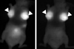
The Rochester Institute of Technology (RIT) and Rochester Regional Health in New York are collaborating to develop thermal breast cancer screening techniques.
Supported by a National Science Foundation grant of $99,620, the two-year project will explore whether modern thermal, or infrared, imaging can serve as an alternative to ultrasound or MRI in a complementary role to conventional mammography.
New thermal imaging techniques can identify alterations in the metabolic behavior of tissues and release specific thermal signatures, according to lead researcher Satish Kandlikar, PhD, from RIT. Thermal imaging will be combined with an artificial intelligence system that predicts the location and size of tumors on a thermal map.
"The team isn't interested in replicating the current methods of thermography; we want to improve the technology enough that thermal imaging will be a fully reliable complement to mammography in breast cancer detection," said Dr. Lori Medeiros, of Rochester Regional Health Breast Center, in a statement.
The project will be conducted under the auspices of the RIT and Rochester Regional Health Alliance, which was established in 2008. RIT is Rochester Regional Health's official academic affiliate, while Rochester Regional is the university's official affiliated clinical partner.