Sunday, November 25 | 12:30 p.m.-1:00 p.m. | LL-BRS-SU5A | Lakeside Learning Center
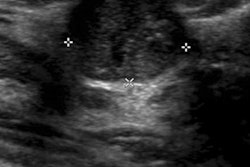
How best can doctors assess residual disease in inflammatory breast cancer (IBC) patients who have undergone neoadjuvant chemotherapy? Find out in this Sunday poster presentation.Researchers from the University of Texas MD Anderson Cancer Center will share findings from a study they conducted to evaluate the performance of mammography, ultrasound, and MRI for this task.
Dr. Sarah Martaindale and colleagues used their institution's IBC Registry protocol to identify women with an inflammatory breast cancer diagnosis who had had baseline and mammography, ultrasound, and MRI exams after neoadjuvant chemotherapy. Between January 2006 and December 2009, 94 women were enrolled in the IBC Registry protocol; 64 patients met the team's inclusion criteria.
They found the following:
- Mean baseline tumor size was 63 mm, 48 mm, and 69 mm, respectively, on mammography, ultrasound, and MRI.
- Breast tumor size on MRI before neoadjuvant chemotherapy was larger than on ultrasound and mammography.
- Tumor size on MRI and ultrasound after neoadjuvant chemotherapy were similar.
- There was a strong association between tumor size after neoadjuvant chemotherapy and higher pathologic response for ultrasound and MRI.
What did the team conclude? While ultrasound and MRI measurement of tumor size after neoadjuvant chemotherapy can predict for residual disease, MRI may be better for monitoring larger tumor size before neoadjuvant chemotherapy.