Sunday, November 25 | 11:05 a.m.-11:15 a.m. | SSA01-03 | Arie Crown Theater
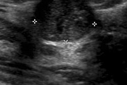
Screening with automated breast ultrasound (ABUS) -- as an adjunct to digital mammography -- detects early-stage, node-negative invasive carcinoma in asymptomatic women with dense breast tissue, according to Dr. Rachel Brem, of George Washington University Medical Center, and colleagues. And finding these cancers could reduce breast cancer mortality rates.In this Sunday morning session, Brem will present findings from a study her team conducted to evaluate the characteristics of cancers in women with more than 50% breast density, identified on automated breast ultrasound with digital mammography.
The study included more than 15,000 asymptomatic women who had no breast surgeries, interventional procedures, or cancer diagnosis and/or treatment in the previous 12 months. In all, 112 cancers were found, with 27% detected only by ABUS (of these, 92% were invasive and 8% were ductal carcinoma in situ).
In addition, 44% of the cancers found only by automated breast ultrasound were grade 1, 44% were grade 2, and 14% were grade 3. Of those invasive cancers identified, 68% were stage IA, 5% were stage IB, 14% were stage IIA, 5% were stage IIB, and 9% were stage IIIA, according to the researchers.
Finding early-stage, node-negative invasive carcinomas in women with dense breasts is likely to affect breast cancer mortality when ABUS is used in combination with digital mammography, compared with digital mammography alone, Brem's team concluded.