Dear Women's Imaging Insider,
Although most radiologists agree that computer-aided detection (CAD) for breast cancer screening helps detect microcalcifications, there's less concord about the technology's ability to identify masses and architectural distortions -- with some radiologists arguing that CAD marks too many false-positive areas.
Is there a way to make CAD more effective? Dutch researchers developed a system that assists radiologists in interpreting suspicious regions rather than helping them detect them, displaying marks "on demand" when readers mouse-click over a particular mammographic area. Find out what they discovered about the pros and cons of "interactive" CAD by clicking here.
We've also covered yesterday's decision by a U.S. Food and Drug Administration panel to recommend approval for a new imaging mode on Hologic's Selenia Dimensions 3D digital breast tomosynthesis system. Click here to read more.
What else is going on in the Women's Imaging Digital Community? Take a look:
- Discover why researchers think breast brachytherapy may produce more complications than benefits for older and elderly women with early-stage breast cancer.
- Check out our coverage of the Minnie's winners, many of which point to the ongoing importance of breast imaging.
- Learn how 3D phase-contrast tomography shows smaller breast tumors.
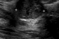
- Read why ultrasound beat mammography as the best imaging modality for initial evaluation of breast cancer in symptomatic women ages 30 to 39.
- Find out how VuCOMP hopes to shake up the mammography CAD market with the premarket approval (PMA) it has received for its M-Vu CAD software for digital mammography.
As always, if you have a comment, report, or article idea to share about any aspect of women's imaging, I invite you to contact me.