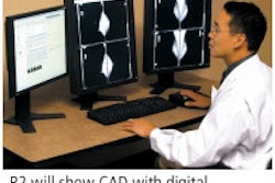
Merge will display a pair of new technologies in its RSNA booth, including an ultrasound CAD workstation and a specialized mammography viewing workstation.
Cedara B-CAD was developed by the Milwaukee company's Cedara Software unit of Mississauga, Ontario. The software automatically analyzes, segments, and classifies shape and orientation characteristics of user-selected regions of interest in breast ultrasound images. It includes annotations based on the ACR BI-RADS Breast Imaging Atlas, and automatically generates reports using the ACR BI-RADS Ultrasound Lexicon Classification Form. The software began shipping this month.
Cedara I-Read Mammo is a dedicated mammography viewing workstation designed to work with images from multiple modalities and vendors. It includes tools such as synchronized magnification and pan and zoom, as well as digital tools that simulate hard-copy review. It can be interfaced with any DICOM-compliant PACS software, according to the company.
By Brian Casey
AuntMinnie.com staff writer
November 7, 2005
Copyright © 2005 AuntMinnie.com

![A normal mammogram confirmed by three-year radiologic follow-up illustrates reader-marked regions of interest (ROIs) during (A) unaided (round 1) and (B) artificial intelligence (AI)–assisted (round 2) reading. Each colored dot represents an ROI for recall by a human reader. Readers could mark more than one ROI per case, represented by multiple dots of the same color. During AI-assisted reading, the AI system displayed three visible prompts: two with suspicion of malignancy scores of 35% (left mediolateral oblique [L MLO] and craniocaudal [L CC]) and one with a suspicion of malignancy score of 10% (right craniocaudal [R CC]), shown as polygonal overlays. Without AI, six of 10 readers (60%) marked a false-positive ROI. With AI assistance, this fell to two of 10 (20%). R MLO = right mediolateral oblique.](https://img.auntminnie.com/mindful/smg/workspaces/default/uploads/2026/07/2026-07-14-radiology-mammogram-ai-auto-bias.H0bYO8QlWs.jpg?auto=format%2Ccompress&fit=crop&h=100&q=70&w=100)


![A normal mammogram confirmed by three-year radiologic follow-up illustrates reader-marked regions of interest (ROIs) during (A) unaided (round 1) and (B) artificial intelligence (AI)–assisted (round 2) reading. Each colored dot represents an ROI for recall by a human reader. Readers could mark more than one ROI per case, represented by multiple dots of the same color. During AI-assisted reading, the AI system displayed three visible prompts: two with suspicion of malignancy scores of 35% (left mediolateral oblique [L MLO] and craniocaudal [L CC]) and one with a suspicion of malignancy score of 10% (right craniocaudal [R CC]), shown as polygonal overlays. Without AI, six of 10 readers (60%) marked a false-positive ROI. With AI assistance, this fell to two of 10 (20%). R MLO = right mediolateral oblique.](https://img.auntminnie.com/mindful/smg/workspaces/default/uploads/2026/07/2026-07-14-radiology-mammogram-ai-auto-bias.H0bYO8QlWs.jpg?auto=format%2Ccompress&fit=crop&h=112&q=70&w=112)