Investigators from the University of Michigan in Ann Arbor are working on an automated spot compression mammography technique that could take the guesswork out of imaging women with dense breasts. Dr. Mitchell Goodsitt from the departments of radiology and environmental health sciences discussed his institution’s work at last year’s RSNA conference.
"Imaging women with dense breast tissue is difficult with screen-film mammography." Goodsitt said. "It’s still a problem even with digital mammography because there’s low signal-to-noise ratio in that region. We’re developing an automated spot mammography technique that involves an automatic detection of large, suspicious, dense regions, auto spot compression, and auto spot collimation."
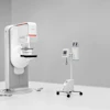
Goodsitt’s group included 10 senior-year mechanical engineering students who designed and built a prototype device that was then attached to a biopsy unit (Fischer Imaging, Denver, CO).
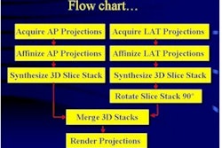
Computerized image analysis was applied to a digital mammogram to segment the dense area and identify large dense regions. The minimum-sized rectangle or ellipse that bounds the dense regions is automatically determined, and the coordinates of the bounding region are then employed to position collimator blades via step motors in a custom-built, asymmetric, and rotatable collimator.
"We take a regular mammogram using a digital system, locate a suspicious region, remove the conventional large compression paddle, restrain the breast with some device, put in a spot compression paddle moving asymmetrically to the suspicious region, and then spot compress and collimate to take a second, more penetrating image just of the suspicious region," he said.
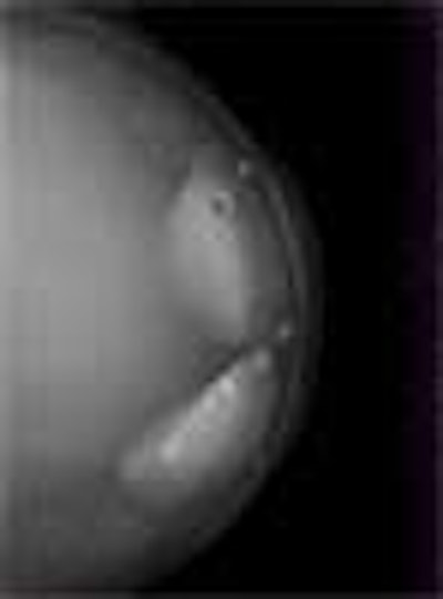
The image at the top was is a full-field image of a compressible breast phantom, acquired with a GE 2000D digital mammography system. Goodsitt said his group plans to develop an automated spot collimation system that can be adapted to this GE unit.

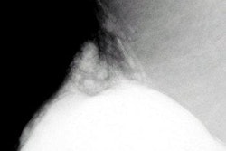
A combined spot compression and spot collimation image obtained on the GE 2000D full-field digital mammography system. Because of the spreading of the tissues, spot collimation, and spot compression, conspicuity of a mass-like object is improved.
The group developed two types of automated software, Goodsitt said. One program allows for a threshold method to segment dense regions. A second program allows the radiologist to use a drawing tool and trace the dense regions. The software also colors the three largest suspicious regions as red, green and blue, he added. Finally, the computer program tells the technologist where to move the device, decreasing the chance of human error.
Goodsitt said his group, along with co-investigator Heang-Ping Chan, would continue to perfect the prototype system, as well as test it on breast phantoms.
By Shalmali PalAuntMinnie.com staff writer
January 30, 2001
Related Reading
Missed breast cancer: Avoiding this pitfall, October 2000
Click here to post your comments about this story. Please include the headline of the article in your message.
Copyright © 2001 AuntMinnie.com
![A normal mammogram confirmed by three-year radiologic follow-up illustrates reader-marked regions of interest (ROIs) during (A) unaided (round 1) and (B) artificial intelligence (AI)–assisted (round 2) reading. Each colored dot represents an ROI for recall by a human reader. Readers could mark more than one ROI per case, represented by multiple dots of the same color. During AI-assisted reading, the AI system displayed three visible prompts: two with suspicion of malignancy scores of 35% (left mediolateral oblique [L MLO] and craniocaudal [L CC]) and one with a suspicion of malignancy score of 10% (right craniocaudal [R CC]), shown as polygonal overlays. Without AI, six of 10 readers (60%) marked a false-positive ROI. With AI assistance, this fell to two of 10 (20%). R MLO = right mediolateral oblique.](https://img.auntminnie.com/mindful/smg/workspaces/default/uploads/2026/07/2026-07-14-radiology-mammogram-ai-auto-bias.H0bYO8QlWs.jpg?auto=format%2Ccompress&fit=crop&h=100&q=70&w=100)


![A normal mammogram confirmed by three-year radiologic follow-up illustrates reader-marked regions of interest (ROIs) during (A) unaided (round 1) and (B) artificial intelligence (AI)–assisted (round 2) reading. Each colored dot represents an ROI for recall by a human reader. Readers could mark more than one ROI per case, represented by multiple dots of the same color. During AI-assisted reading, the AI system displayed three visible prompts: two with suspicion of malignancy scores of 35% (left mediolateral oblique [L MLO] and craniocaudal [L CC]) and one with a suspicion of malignancy score of 10% (right craniocaudal [R CC]), shown as polygonal overlays. Without AI, six of 10 readers (60%) marked a false-positive ROI. With AI assistance, this fell to two of 10 (20%). R MLO = right mediolateral oblique.](https://img.auntminnie.com/mindful/smg/workspaces/default/uploads/2026/07/2026-07-14-radiology-mammogram-ai-auto-bias.H0bYO8QlWs.jpg?auto=format%2Ccompress&dpr=2&fit=crop&h=167&q=70&w=250)


