
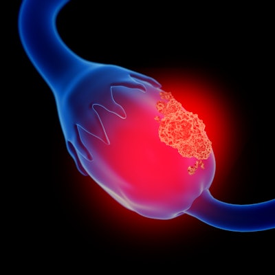
O-RADS MRI scoring is an effective way to assess malignancy risk in adnexal masses referred after inconclusive ultrasound results, but improvements are needed for the metric, according to a commentary published May 13 in European Radiology.
A group led by Dr. Isabelle Thomassin-Naggara, PhD, of the Hôpital Tenon in Paris wrote that additional MR criteria may be added to the scoring metric to better find borderline cystadenomas among malignant tumors, and that current trials may validate the method to inform clinical management of patients with potential ovarian cancer.
"O-RADS MRI score is a robust, reproducible, and simple way to assess the risk of malignancy of an adnexal mass referred after ultrasonography," Thomassin-Naggara and co-authors noted.
O-RADS MRI is a scoring system that characterizes adnexal masses found on imaging and assessing risk. A 2022 study by researchers in France and the U.K., led by Thomassin-Naggara and colleagues, found that their five-point scoring system with MRI could distinguish between malignant and benign ovarian cysts with an accuracy of 90%.
The authors wrote that the scoring system's better specificity can reclassify masses as benign, indeterminate, or malignant on ultrasonography.
Thomassin-Naggara's team wrote that there are two next steps to improve the use of this scoring method.
The first is to include O-RADS MRI scoring into clinical management guidelines, which would have a significant impact during multidisciplinary tumor board sessions. In fact, two ongoing studies are focusing on this: One is the Ascordia study in France, which is exploring the impact of an MR scoring system of therapeutic strategy of pelvic adnexal masses, and the other is the MR in ovarian cancer (MROC) study in the U.K., which is evaluating the ability of O-RADS MRI scoring to identify the impact of the score on treatment planning.
The second is having clinicians better define the type of malignant tumors found, the authors wrote.
"This is particularly relevant in premenopausal women in whom the surgeon may opt for a conservative surgery if the tumor is only a serous borderline cystadenoma," they noted. "Hence, some researchers evaluated additional MR criteria to improve O-RADS MRI score and particularly O-RADS MRI 4 category, which is the less informative category due to its large positive predictive value range."


![Overview of the study design. (A) The fully automated deep learning framework was developed to estimate body composition (BC) (defined as subcutaneous adipose tissue [SAT] in liters; visceral adipose tissue [VAT] in liters; skeletal muscle [SM] in liters; SM fat fraction [SMFF] as a percentage; and intramuscular adipose tissue [IMAT] in deciliters) from MRI. The fully automated framework comprised one model (model 1) to quantify different BC measures (SAT, VAT, SM, SMFF, and IMAT) as three-dimensional (3D) measures from whole-body MRI scans. The second model (model 2) was trained to identify standardized anatomic landmarks along the craniocaudal body axis (z coordinate field), which allowed for subdividing the whole-body measures into different subregions typically examined on clinical routine MRI scans (chest, abdomen, and pelvis). (B) BC was quantified from whole-body MRI in over 66,000 individuals from two large population-based cohort studies, the UK Biobank (UKB) (36,317 individuals) and the German National Cohort (NAKO) (30,291 individuals). Bar graphs show age distribution by sex and cohort. BMI = body mass index. (C) After the performance assessment of the fully automated framework, the change in BC measures, distributions, and profiles across age decades were investigated. Age-, sex-, and height-adjusted body composition reference curves were calculated and made publicly available in a web-based z-score calculator (https://circ-ml.github.io).](https://img.auntminnie.com/mindful/smg/workspaces/default/uploads/2026/05/body-comp.XgAjTfPj1W.jpg?auto=format%2Ccompress&fit=crop&h=100&q=70&w=100)


![Overview of the study design. (A) The fully automated deep learning framework was developed to estimate body composition (BC) (defined as subcutaneous adipose tissue [SAT] in liters; visceral adipose tissue [VAT] in liters; skeletal muscle [SM] in liters; SM fat fraction [SMFF] as a percentage; and intramuscular adipose tissue [IMAT] in deciliters) from MRI. The fully automated framework comprised one model (model 1) to quantify different BC measures (SAT, VAT, SM, SMFF, and IMAT) as three-dimensional (3D) measures from whole-body MRI scans. The second model (model 2) was trained to identify standardized anatomic landmarks along the craniocaudal body axis (z coordinate field), which allowed for subdividing the whole-body measures into different subregions typically examined on clinical routine MRI scans (chest, abdomen, and pelvis). (B) BC was quantified from whole-body MRI in over 66,000 individuals from two large population-based cohort studies, the UK Biobank (UKB) (36,317 individuals) and the German National Cohort (NAKO) (30,291 individuals). Bar graphs show age distribution by sex and cohort. BMI = body mass index. (C) After the performance assessment of the fully automated framework, the change in BC measures, distributions, and profiles across age decades were investigated. Age-, sex-, and height-adjusted body composition reference curves were calculated and made publicly available in a web-based z-score calculator (https://circ-ml.github.io).](https://img.auntminnie.com/mindful/smg/workspaces/default/uploads/2026/05/body-comp.XgAjTfPj1W.jpg?auto=format%2Ccompress&fit=crop&h=112&q=70&w=112)


