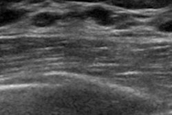
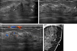
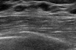
Sonographic imaging descriptors and histopathological diagnoses are needed for more accurate assessment of benign breast lesions, suggest findings published February 3 in BMC Women’s Health.
Researchers led by Anwar Rjoop, MD, from the Jordan University of Science and Technology in Irbin, Jordan, found that the presence of an oval-shaped mass and posterior shadowing are significant imaging markers for assessing benign breast lesions categorized as BI-RADS 4c or 5.
“The study recommends a correlation between clinical and radiological findings and encourages multidisciplinary decision-making among radiologists, pathologists, and clinicians to determine if a repeat biopsy is warranted,” Rjoop and colleagues wrote.
BI-RADS 4c and 5 categories indicate strong to certain malignancy for breast cancer and prompt biopsy. Still, this system is imperfect, with imaging findings of some benign breast lesions mimicking malignant characteristics.
The investigators identified histopathological diagnosis trends of benign breast masses classified into these BI-RADS subgroups. They also studied the radiological characteristics of these masses and identified ultrasound features that could lead to false-positive results.
The team reviewed breast lesions reported as BI-RADS 4c or 5 on breast ultrasound that underwent either ultrasound-guided core needle or stereotactic vacuum-assisted biopsy. It also compared imaging descriptors to those of a matched control of 50 malignant cases with BI-RADS 4c.
The retrospective single-facility study included data collected between 2015 and 2022 from 828 breast lesions with the aforementioned BI-RADS categories from the same number of women.
Of the total lesions, 44 (5.3%) were deemed benign at initial biopsy while 784 lesions (94.7%) were malignant. After histopathological testing and repeat biopsy, 26 women (3.1%) had a discordant benign diagnosis. Half of the repeated biopsies (10 of 20) showed malignant pathology.
Compared with imaging findings in the control group, the presence of an oval-shaped mass was significantly more common in women with benign pathology (p = 0.035). However, the presence of posterior shadowing was significantly less common (p = 0.05) in benign lesions. The researchers observed no significant differences in other imaging characteristics.
Finally, the most common histopathological diagnosis was fibrocystic change, the team noted.
The study authors highlighted the potential of sonoelastography and AI in aiding with decision-making to end unnecessary procedures such as biopsy. They also highlighted the need for continuous research to improve the diagnosis and treatment of breast lesions and reduce false-positive rates by incorporating other methodologies.
“It [the study] suggests reducing false-positive rates and unnecessary biopsies in benign lesions and establishing clinical guidelines for repeating biopsy in patients with high-risk radiological features,” they added.
The full study can be found here.