Dear Women's Imaging Insider,
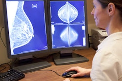
Digital breast tomosynthesis (DBT) has been shown in clinical studies to reduce recalls and boost cancer detection rates -- across all tissue densities. But when used in combination with 2D full-field digital mammography, it imparts additional radiation dose. So some DBT vendors have developed software that synthesizes 2D images from the tomosynthesis data, thereby reducing women's radiation exposure.
How do these synthesized images compare to traditional digital mammography? Just fine, according to researchers from the University of Utah in Salt Lake City. A team led by Dr. Phoebe Freer found that synthesized 2D plus DBT can replace digital mammography plus DBT without concern that performance measures will be negatively affected. Learn more in our Insider Exclusive.
After you've read our featured article, take a look at what else is going on in our Women's Imaging Community:
- Find out what South Korean researchers have to say about the prognosis for women whose breast cancers are detected on screening ultrasound -- and factors that could contribute to recurrence.
- Learn how one academic practice reduced mammography recall rates with a few simple interventions.
- Are older women getting appropriate breast cancer screening? Perhaps not, say researchers from Dana-Farber Cancer Institute in Boston.
- Read why radiologists can trust DBT breast density measurements.
- Discover how machine learning can help predict pregnancies with fetal growth restriction -- and how this information could lead to interventions that would protect vulnerable fetuses.
And of course, if you have a comment, report, or article idea to share about any aspect of women's imaging, please contact me.