Dear AuntMinnie Member,
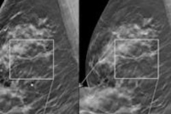
Digital breast tomosynthesis (DBT) is an exciting new technology that has the potential to revolutionize breast screening. But it's being implemented at uneven rates around the U.S., according to a new article in our Women's Imaging Digital Community.
Researchers from the University of Colorado at Denver performed a survey to analyze the use of DBT and which patients are being selected to receive it. They found that while tomosynthesis is becoming more common, it's still a limited resource. Learn more by clicking here.
In other news in the community, check out this article on how radiologists who knew about women's risk of breast cancer turned in a better diagnostic performance in reading screening mammograms than those who read images without risk information.
And make sure to read this story on how radiologists reading synthesized 2D images reconstructed from 3D tomosynthesis data had equivalent diagnostic performance -- at a lower radiation dose -- compared to readers interpreting a combination of DBT and full-field digital mammography exams.
Big data and personalized care
We're highlighting a couple of intriguing new articles in our Imaging Informatics Community, including a presentation by Dr. Daniel Rubin from Stanford University at last week's International Symposium on Multidetector-Row CT in San Francisco.
Dr. Rubin believes we are seeing the development of rapid-learning systems, which can mine vast amounts of data in historical archives to guide better decision-making in treatment. This will result in true evidence-based care, he believes. Read more by clicking here.
Also check out this article on what to look for when contemplating the purchase of a universal viewer, the front-end piece of the puzzle that enables physicians throughout the healthcare enterprise to view medical images stored on vendor-neutral archives.
Dodging a MedPAC bullet?
Finally, did radiology just dodge a bullet when it comes to new reimbursement cuts? The Medicare Payment Advisory Commission (MedPAC) last week issued its new report to the U.S. Congress on revising Medicare payment policies, and (fortunately) the report contained no new recommendations for medical imaging. See for yourself by clicking here.