Dear AuntMinnie Member,
Mammography is a discipline beset by challenges, ranging from low reimbursement to medicolegal liability to regulatory burdens. Even one new technology first thought to be mammography's savior -- computer-aided detection -- has been besmirched by a recent study questioning its effectiveness.
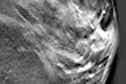
Riding to the rescue is digital breast tomosynthesis (DBT), a new technology based on full-field digital mammography that uses a gantry head that pans across the breast to collect 2D slices that can be reconstructed into 3D volumes. Developers of DBT systems believe the technology can help breast imagers visualize pathology that might have been obscured by overlapping tissue on 2D images.
But even when the first DBT system gets to market, there could be major impediments to widespread adoption -- in spite of the technology's clinical benefits. Find out what some of the barriers could be in a new article in our Women's Imaging Digital Community, which you can reach by clicking here.
In another new article in the community, research based on data from Sweden indicates that mammography screening can significantly reduce the rates of large tumor and lymph node-positive breast cancer. That article is available by clicking here.
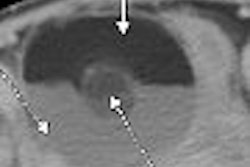
Finally, we're pleased to highlight a case study out of Africa that demonstrates the use of CT to detect an ovarian dermoid cyst. Get the details of the case by clicking here.
You'll find these stories and more in our Women's Imaging Digital Community, at women.auntminnie.com.