Dear Women's Imaging Insider,
Women's imaging is truly a multimodality discipline, as evidenced by this installment of the Insider.
First, we have good news and not so good news about breast biopsy. Radiologists in New York City have achieved success with MRI-guided biopsy for women at high risk of breast cancer or with problematic mammograms. Their clinical experience bolsters the value of image-guided breast biopsy on the whole. Unfortunately, this information is not necessarily being taken into consideration by referring physicians, according to a paper by a group from Brown University in Providence, RI. To read more in the Insider Exclusive, click here.
In the Women's Imaging Digital Community, you'll also find a report from St. Louis' Mallinckrodt Institute of Radiology on the role of FDG-PET in predicting outcomes after cervical cancer treatment.
In ultrasound news, there's word that sonography can play a role in detecting residual primary breast malignancy in patients receiving neoadjuvant chemotherapy.
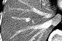
On the CT front, researchers from the Memorial Sloan-Kettering Cancer Center in New York City discuss using the modality most effectively for finding hepatic lesions in women with breast cancer.
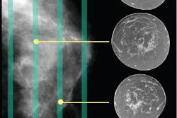
Finally, check out an article on full-field digital mammography and the challenges it presents for image management.