AI could help standardize the acquisition of shoulder ultrasound images, according to a study published March 27 in Academic Radiology.
A team led by Yanni He, PhD, from the Affiliated Guangdong Second Provincial General Hospital of Jinan University in Guangzhou, China, developed an AI algorithm and reported its success in providing accurate, real-time guidance.
“[The AI system] reduces operator dependency for novices and establishes a reproducible foundation for subsequent diagnostic interpretation, representing a critical advancement towards standardized musculoskeletal [MSK] ultrasound imaging acquisition,” the He team wrote.
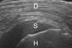
While ultrasound has diagnostic value in MSK imaging, even experienced MSK radiologists may face challenges in image interpretation. This is due to the complex anatomy of the shoulder, including similar tendons surrounding the humerus within the shoulder.
Previous research suggests that AI can help reduce user dependence during ultrasound exams.
He and colleagues developed an AI-guided algorithm for real-time automatic classification and structural recognition of shoulder ultrasound planes. Their goal was to standardize the ultrasound scanning process.
The team used 852 standard plane images and 74,909 frame images from 13,312 shoulder ultrasound exams from a single center for model training and internal testing. For the external test set, it used 8,458 frame images from 480 videos collected from a second center. From there, the researchers developed convolutional neural networks (CNNs).
The resulting multitask AI system (EfficientNetB2) can concurrently guide image acquisition of 15 standard planes and localize 27 key structures, the researchers reported. The study included 137 adult volunteers, with 98 in the training set, 23 in the internal test set, and 16 in the external test set.
The system achieved an area under the curve (AUC) of 0.99 and a mean average precision of 0.89 in an independent external validation set. It also achieved an average accuracy of 94% and F1 scores ranging from 0.87 to 0.99.
For junior residents, AI guidance significantly reduced shoulder ultrasound exam time by 34% compared to unassisted scans (10.1 minutes versus 15.3 minutes; p = 0.014).
“Independent expert evaluation confirmed the high real-time guidance accuracy of the system,” the researchers wrote.
The study authors also suggested that this system could have applications in tele-ultrasound and patient self-monitoring.
“The system provides a promising training tool and a foundation for consistent imaging,” they added.
The team called for more studies to determine whether this AI-guided acquisition leads to improved diagnostic accuracy or clinical decision-making.
Read the full study here.