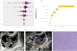
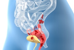
Deep infiltrating endometriosis on transvaginal ultrasound (TVUS) decreases the chance for a live birth in women undergoing their first fertility treatment, according to Swedish research published January 19 in Fertility and Sterility.
A team led by Sara Alson, MD, from Lund University in Malmö found that expectant mothers undergoing in vitro fertilization (IVF) or intracytoplasmic sperm injection (ICSI) treatment and who have endometriosis had a nearly 10% lower chance of a live birth compared with those without the disease.
“Our results contradict those that suggest that IVF treatment can completely counter the negative effects of endometriosis seen on fertilization in vivo,” Alson and co-authors wrote.
Endometriosis affects about 10-15% of women of reproductive age and is tied to chronic inflammation and subfertility. However, the cause of subfertility by endometriosis is not well understood.
IVF and ICSI are assisted reproductive treatments explored as potential ways to counteract the subfertility effects of endometriosis. There is conflicting evidence, though, as to their respective effectiveness on liver birth rates. TVUS meanwhile is a first-line imaging approach to diagnose endometriomas and deep infiltrating endometriosis.
The Alson team evaluated the cumulative live birth rate after the first IVF or ICSI treatment in women with or without deep infiltrating endometriosis diagnosed by TVUS. It used group definitions from the International Deep Endometriosis Analysis.
In their prospective study, the researchers included data from 1,040 subfertile women ages 25 to 39. These women underwent their first IVF or ICSI treatment between 2019 and 2022. Of the total, 234 women were diagnosed with deep infiltrating endometriosis through TVUS before fertility treatment began.
The cumulative live birth rate after the first fertility treatment regimen in the study population was 41%.
The team reported that women diagnosed with deep infiltrating endometriosis had a cumulative live birth rate of 33.3% (n = 78/234). Women without the disease had a rate of 43.2% (n = 348/806, p = 0.007).
Additionally, the cumulative live birth rate for women with deep infiltrating endometriosis had a crude relative risk of 0.77. After adjustments were made for patient characteristics such as age, body mass index (BMI), s-anti Müllerian hormone, stimulation protocol, and day for embryo transfer, the adjusted relative risk was 0.63.
Finally, the team reported no significant difference in the number of retrieved mature oocytes, fertilization rate, or good-quality embryos between women with and without endometriosis.
The study authors suggested multiple reasons why their results contradict previous studies in this area. For example, prior studies were retrospective and used different methods in diagnosing endometriosis.
The authors also called for future research to find out whether women with deep infiltrating endometriosis could achieve the same cumulative live birth rate as women without the disease after repeated cycles of fertility treatment.
“Future studies will also reveal whether a concurrent presence of adenomyosis diagnosed with TVUS affects the IVF/ICSI outcome,” they added.
The full study can be found here.

![Examples of ultrasound findings and techniques. (A) Images in a 39-year-old male patient with a mass in the left thigh. The mass is heterogeneous on the B-mode US image (compared with the patient in D) and showed increased microvascularity (superb microvascular imaging [SMI]) and shear-wave elastography (SWE) values. Undifferentiated pleomorphic sarcoma was diagnosed at biopsy (with pleomorphic rhabdomyosarcoma in surgical specimen). (B) Images in an 18-year-old male patient with a mass in the left leg. The mass is hypoechoic on the B-mode image, with no other findings suggestive of malignancy. The lesion is in contact with the cortex of the tibia, which is slightly irregular. CT revealed a doubtful anteromedial tibial erosion. The microvascular study demonstrated high vascularization, suggestive of malignancy. Periosteal Ewing sarcoma was diagnosed with both histologic and immunohistochemical confirmation. (C) Images in a 69-year-old female patient with a lump growing on the outside of the left leg. Multiple SWE examinations were performed (please note the high values obtained in the measurements, whereas the color map highlights the stiffness relative to adjacent tissues). SMI showed areas of increased vascularization to target for sampling. Undifferentiated spindle cell sarcoma was diagnosed at biopsy, with residual leiomyosarcoma in the surgical specimen after neoadjuvant therapy. (D) Images in a 56-year-old female patient with a mass in the right thigh. The mass is heterogeneous at both B-mode ultrasound (similar to patient A) and MRI (coronal T2-weighted spectral attenuated inversion recovery [SPAIR]; T1-weighted pre-contrast and postcontrast imaging), which even shows uptake after the administration of paramagnetic contrast material, which is traditionally suggestive of malignancy. Low values at SMI and elastography are suggestive of benignity. Spindle cell lipoma was diagnosed at biopsy, with atypical spindle cell lipomatous tumor in the surgical specimen.](https://img.auntminnie.com/mindful/smg/workspaces/default/uploads/2026/08/images-radiol250278fig2.APCFLSvX6p.jpg?auto=format%2Ccompress&fit=crop&h=100&q=70&w=100)


![Examples of ultrasound findings and techniques. (A) Images in a 39-year-old male patient with a mass in the left thigh. The mass is heterogeneous on the B-mode US image (compared with the patient in D) and showed increased microvascularity (superb microvascular imaging [SMI]) and shear-wave elastography (SWE) values. Undifferentiated pleomorphic sarcoma was diagnosed at biopsy (with pleomorphic rhabdomyosarcoma in surgical specimen). (B) Images in an 18-year-old male patient with a mass in the left leg. The mass is hypoechoic on the B-mode image, with no other findings suggestive of malignancy. The lesion is in contact with the cortex of the tibia, which is slightly irregular. CT revealed a doubtful anteromedial tibial erosion. The microvascular study demonstrated high vascularization, suggestive of malignancy. Periosteal Ewing sarcoma was diagnosed with both histologic and immunohistochemical confirmation. (C) Images in a 69-year-old female patient with a lump growing on the outside of the left leg. Multiple SWE examinations were performed (please note the high values obtained in the measurements, whereas the color map highlights the stiffness relative to adjacent tissues). SMI showed areas of increased vascularization to target for sampling. Undifferentiated spindle cell sarcoma was diagnosed at biopsy, with residual leiomyosarcoma in the surgical specimen after neoadjuvant therapy. (D) Images in a 56-year-old female patient with a mass in the right thigh. The mass is heterogeneous at both B-mode ultrasound (similar to patient A) and MRI (coronal T2-weighted spectral attenuated inversion recovery [SPAIR]; T1-weighted pre-contrast and postcontrast imaging), which even shows uptake after the administration of paramagnetic contrast material, which is traditionally suggestive of malignancy. Low values at SMI and elastography are suggestive of benignity. Spindle cell lipoma was diagnosed at biopsy, with atypical spindle cell lipomatous tumor in the surgical specimen.](https://img.auntminnie.com/mindful/smg/workspaces/default/uploads/2026/08/images-radiol250278fig2.APCFLSvX6p.jpg?auto=format%2Ccompress&fit=crop&h=112&q=70&w=112)