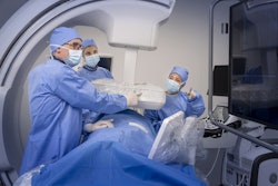
GE HealthCare (GEHC) and University of California (UC) San Diego Health have entered a strategic enterprise collaboration that will bring medical imaging technologies to the latter.
The collaboration's goal is to integrate imaging and medical technologies throughout the UC San Diego Health system for improved outcomes and greater cross-functional connectivity. This includes leveraging emerging modalities and technologies such as theranostics and AI for care delivery and streamlining workflows.
GEHC will also offer technical service support for clinician education and a service delivery model to help support preventative maintenance for connected equipment.
Some GEHC imaging products that will be implemented include the StarGuide SPECT/CT system, Smart Subscription for CT, and the Signa Continuum program for MR.
In other GEHC news, the company has launched its latest AI-enabled Venue family of point-of-care ultrasound (POCUS) systems.
New AI-enabled tools extend across the entire Venue portfolio, including Venue, Venue Go, Venue Fit, and Venue Sprint systems, and include the following:
Nerveblox, a tool that automatically labels anatomical landmarks to assist clinicians in performing 12 of the most common peripheral nerve blocks.
Auto Bladder Volume, which simplifies urinary bladder volume measurement from two views. This tool is also available on the wireless Vscan Air CL probe, GE noted.
Contrast Enhanced Ultrasound, which enables dynamic bedside evaluation of abdominal injuries and lesions.
Automated Function Imaging, which provides objective analysis of myocardial strain across heart chambers.
The new tools are designed to simplify complex exams, enhance speed and accuracy during assessments, and support clinicians across all experience levels in delivering confident care, according to the firm.