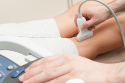
Omitting physical exams before ultrasound exams does not affect diagnostic outcomes, according to findings published September 18 in the European Journal of Radiology.
A team led by Thomas Kwee, MD, PhD, from University Medical Center Groningen in the Netherlands and colleagues reported that more than half of referrals for diagnostic ultrasound skipped physical exams. And while omitting physical exams initially led to reduced clinical reasoning and diagnostic yield, these associations did not remain significant on multivariate analysis.
“Radiologists should therefore adjust their expectation that physical examination prior to imaging can be taken for granted,” the Kwee team wrote.
A traditional part of medical diagnosis, the quality of physical exams has declined over the years. The researchers pointed to several factors for this trend, including the availability of advanced diagnostic technologies, defensive medical practices that rely more on technology-based diagnosis, and not enough emphasis on physical exams in medical curricula, among others.
Debate continues as to whether ultrasound can be a standalone diagnostic technique or should serve as an extension of the physical exam.
Kwee and colleagues explored trends on how often physicians perform physical exams prior to referring patients for diagnostic ultrasound. They also studied whether omitting physical exams affects clinical reasoning quality or diagnostic yield.
The study included 302 patients with a median age of 56 years. The researchers asked patients who underwent diagnostic ultrasound exams whether their referring physician had performed a physical exam. They also defined clinical reasoning quality as the alignment between clinically suspected and ultrasound findings.
The team reported that the physical exam was omitted in 168 cases (55.6%). It also found that omitting the physical exam was less likely in referrals from the following: surgical specialties (odds ratio [OR], 0.490, p = 0.032), family medicine (OR, 0.253, p = 0.016), and residents (OR, 0.303, p < 0.001), cases of new complaints (OR, 0.226, p < 0.001), and head and neck ultrasound (OR, 0.261, p < 0.001).
On univariate analysis, omitting the exam led to marginally lower clinical reasoning scores (p = 0.05), but this trend did not remain after adjusting for the following variables: patient age and gender, the requesting medical specialty, the healthcare professional who requested the ultrasound, main indication for ultrasound, and anatomical region of interest.
Of all ultrasound exams, 51% were positive. Omitting the physical exam showed a trend toward lower diagnostic yield (p = 0.059), but this did not remain significant after adjustment. On multivariate analysis, only ultrasound exams that were requested for surveillance imaging (OR, 0.357, p = 0.004) and ultrasound exams of the head and neck region (OR, 0.392, p = 0.004) were significantly less likely to have a positive result.
The study authors offered two possible explanations for their findings. One is that the diagnostic utility of physical exams may be overestimated in some clinical contexts. The other is the possibility that the overall quality of the physical exam as performed by referring physicians does not reach the expected standard.
“As reliance on imaging increases, our findings raise important considerations for medical training and workflow distribution, signaling a shift in diagnostic responsibility toward imaging departments when clinical assessment is abbreviated or absent,” the authors wrote. “Policymakers should respond to this development by reinforcing physical examination training and ensuring radiology services are adequately resourced to meet increased demand.”
Read the full study here.