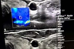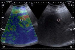
Shear-wave elastography values are higher in children with Hashimoto's thyroiditis and Graves' disease compared with healthy children, a Turkish study published December 9 in Archives de Pédiatrie has found.
Although the study findings are promising for identifying autoimmune thyroid diseases in children, authors Dr. Eda Celebi Bitkin and Dr. Nursen Toprak from Van Yüzüncü Yıl University wrote, although they noted that elastography by itself is not enough to differentiate between the two diseases.
"If the clinical aspect is poor or thyroid-stimulating hormone receptor antibodies are missing, the combined use of conventional ultrasound and acoustic radiation force impulse may be helpful in distinguishing between the two diseases," the authors wrote.
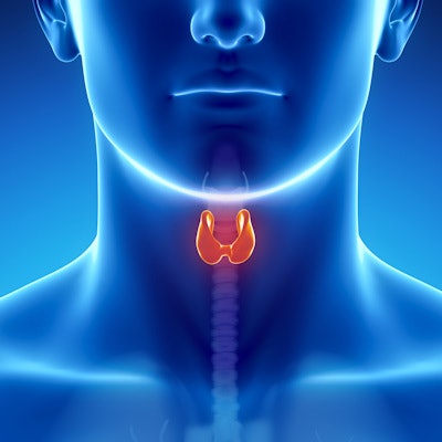
Hashimoto's thyroiditis and Graves' disease are autoimmune diseases that affect the thyroid gland. Ultrasound findings and autoantibody elevation are typically used to diagnose either disease. In rare cases, ultrasound findings may be normal in both diseases, but previous research has suggested that shear-wave velocity values from elastography are higher in autoimmune thyroiditis than in healthy thyroid glands.
Bitkin and Toprak explored the use of elastography in 88 pediatric patients with Hashimoto's thyroiditis and Graves' disease. Out of these, 28 had Hashimoto's thyroiditis, 20 had Graves' disease, and the remaining 40 were healthy controls. All patients underwent thyroid elastography and ultrasound.
The duo found that shear-wave velocity values of patients with either disease showed higher thyroid tissue stiffness than healthy controls, but when such values were compared between both diseases, the team found no significant difference (p = 0.73). As for conventional ultrasound, patients with Hashimoto's thyroiditis had a hypoechoic echo pattern similar to those with Graves' disease (p < 0.05).
"These ultrasound imaging findings reflect longer-term inflammation and may not be found in the early stages of thyroiditis," Bitkin and Toprak wrote.
The researchers did not track disease stage in their study but suggested that the degree of tissue stiffness may change as the diseases progress. They called for future studies to consider disease staging, especially for patients with Hashimoto's thyroiditis.


![Examples of ultrasound findings and techniques. (A) Images in a 39-year-old male patient with a mass in the left thigh. The mass is heterogeneous on the B-mode US image (compared with the patient in D) and showed increased microvascularity (superb microvascular imaging [SMI]) and shear-wave elastography (SWE) values. Undifferentiated pleomorphic sarcoma was diagnosed at biopsy (with pleomorphic rhabdomyosarcoma in surgical specimen). (B) Images in an 18-year-old male patient with a mass in the left leg. The mass is hypoechoic on the B-mode image, with no other findings suggestive of malignancy. The lesion is in contact with the cortex of the tibia, which is slightly irregular. CT revealed a doubtful anteromedial tibial erosion. The microvascular study demonstrated high vascularization, suggestive of malignancy. Periosteal Ewing sarcoma was diagnosed with both histologic and immunohistochemical confirmation. (C) Images in a 69-year-old female patient with a lump growing on the outside of the left leg. Multiple SWE examinations were performed (please note the high values obtained in the measurements, whereas the color map highlights the stiffness relative to adjacent tissues). SMI showed areas of increased vascularization to target for sampling. Undifferentiated spindle cell sarcoma was diagnosed at biopsy, with residual leiomyosarcoma in the surgical specimen after neoadjuvant therapy. (D) Images in a 56-year-old female patient with a mass in the right thigh. The mass is heterogeneous at both B-mode ultrasound (similar to patient A) and MRI (coronal T2-weighted spectral attenuated inversion recovery [SPAIR]; T1-weighted pre-contrast and postcontrast imaging), which even shows uptake after the administration of paramagnetic contrast material, which is traditionally suggestive of malignancy. Low values at SMI and elastography are suggestive of benignity. Spindle cell lipoma was diagnosed at biopsy, with atypical spindle cell lipomatous tumor in the surgical specimen.](https://img.auntminnie.com/mindful/smg/workspaces/default/uploads/2026/08/images-radiol250278fig2.APCFLSvX6p.jpg?auto=format%2Ccompress&fit=crop&h=100&q=70&w=100)


![Examples of ultrasound findings and techniques. (A) Images in a 39-year-old male patient with a mass in the left thigh. The mass is heterogeneous on the B-mode US image (compared with the patient in D) and showed increased microvascularity (superb microvascular imaging [SMI]) and shear-wave elastography (SWE) values. Undifferentiated pleomorphic sarcoma was diagnosed at biopsy (with pleomorphic rhabdomyosarcoma in surgical specimen). (B) Images in an 18-year-old male patient with a mass in the left leg. The mass is hypoechoic on the B-mode image, with no other findings suggestive of malignancy. The lesion is in contact with the cortex of the tibia, which is slightly irregular. CT revealed a doubtful anteromedial tibial erosion. The microvascular study demonstrated high vascularization, suggestive of malignancy. Periosteal Ewing sarcoma was diagnosed with both histologic and immunohistochemical confirmation. (C) Images in a 69-year-old female patient with a lump growing on the outside of the left leg. Multiple SWE examinations were performed (please note the high values obtained in the measurements, whereas the color map highlights the stiffness relative to adjacent tissues). SMI showed areas of increased vascularization to target for sampling. Undifferentiated spindle cell sarcoma was diagnosed at biopsy, with residual leiomyosarcoma in the surgical specimen after neoadjuvant therapy. (D) Images in a 56-year-old female patient with a mass in the right thigh. The mass is heterogeneous at both B-mode ultrasound (similar to patient A) and MRI (coronal T2-weighted spectral attenuated inversion recovery [SPAIR]; T1-weighted pre-contrast and postcontrast imaging), which even shows uptake after the administration of paramagnetic contrast material, which is traditionally suggestive of malignancy. Low values at SMI and elastography are suggestive of benignity. Spindle cell lipoma was diagnosed at biopsy, with atypical spindle cell lipomatous tumor in the surgical specimen.](https://img.auntminnie.com/mindful/smg/workspaces/default/uploads/2026/08/images-radiol250278fig2.APCFLSvX6p.jpg?auto=format%2Ccompress&fit=crop&h=112&q=70&w=112)